

Macrophage endocytic trafficking of antiretroviral nanoparticles
- PMID: 21417829
- PMCID: PMC3184214
- DOI: 10.2217/nnm.11.27
Macrophage endocytic trafficking of antiretroviral nanoparticles
Abstract
Aim: Nanoformulated antiretroviral therapy can improve drug compliance for people infected with HIV. Additional benefits would include specific drug deliveries to viral reservoirs and reduction in systemic toxicities.
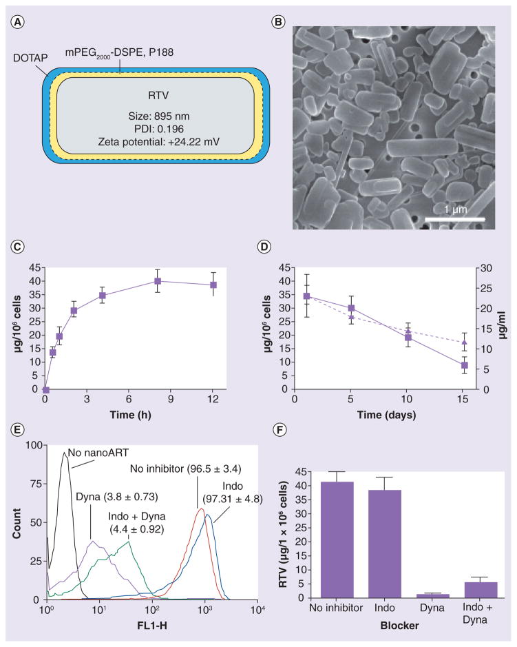
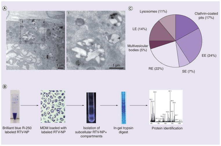
Methods: In this article, we describe mechanisms of crystalline antiretroviral nanoparticle (NP) uptake, intracellular trafficking and release in human monocyte-derived macrophages.
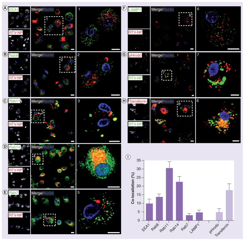
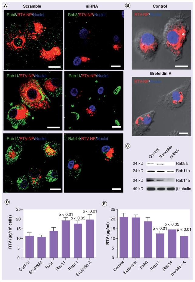
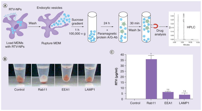
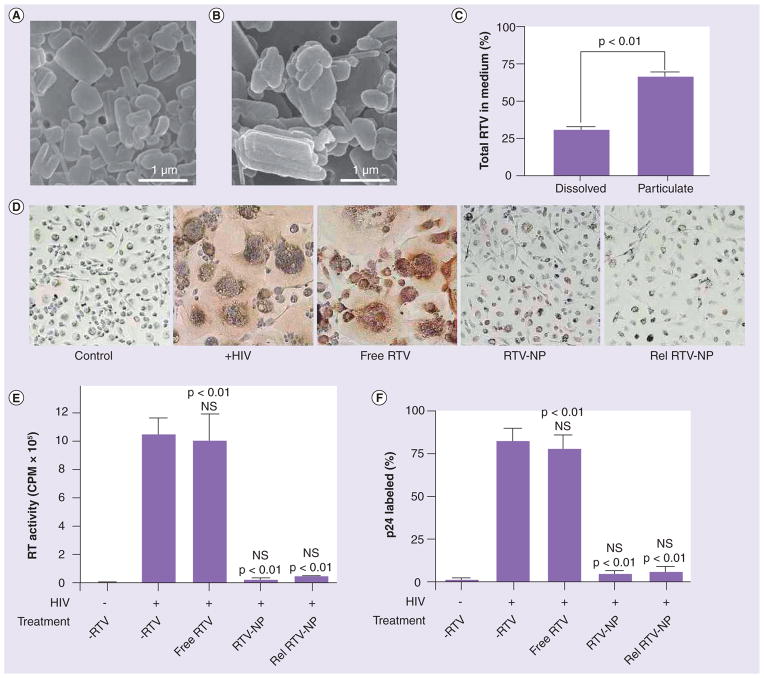
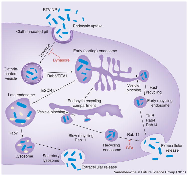
Results: Following clathrin-dependent endocytosis NPs bypassed lysosomal degradation by sorting from early endosomes to recycling endosome pathways. Disruption of this pathway by siRNAs or brefeldin-A impaired particle release. Proteomic and biological analysis demonstrated that particle recycling was primarily Rab11 regulated. Particles were released intact and retained complete antiretroviral efficacy.
Conclusion: These results suggest possible pathways of subcellular transport of antiretroviral nanoformulations that preserve both particle integrity and antiretroviral activities demonstrating the potential utility of this approach for targeted drug delivery.
Figures
Similar articles
-
Efficacy of Tat-conjugated ritonavir-loaded nanoparticles in reducing HIV-1 replication in monocyte-derived macrophages and cytocompatibility with macrophages and human neurons.AIDS Res Hum Retroviruses. 2011 Aug;27(8):853-62. doi: 10.1089/AID.2010.0295. Epub 2011 Feb 2. AIDS Res Hum Retroviruses. 2011. PMID: 21175357 Free PMC article.
-
Endosomal trafficking of nanoformulated antiretroviral therapy facilitates drug particle carriage and HIV clearance.J Virol. 2014 Sep 1;88(17):9504-13. doi: 10.1128/JVI.01557-14. Epub 2014 Jun 11. J Virol. 2014. PMID: 24920821 Free PMC article.
-
Analyses of nanoformulated antiretroviral drug charge, size, shape and content for uptake, drug release and antiviral activities in human monocyte-derived macrophages.J Control Release. 2011 Mar 10;150(2):204-11. doi: 10.1016/j.jconrel.2010.11.019. Epub 2010 Nov 23. J Control Release. 2011. PMID: 21108978 Free PMC article.
-
Lopinavir/ritonavir: a review of its use in the management of HIV infection.Drugs. 2003;63(8):769-802. doi: 10.2165/00003495-200363080-00004. Drugs. 2003. PMID: 12662125 Review.
-
Lopinavir-Ritonavir: a new protease inhibitor.Pharmacotherapy. 2001 Nov;21(11):1352-63. doi: 10.1592/phco.21.17.1352.34419. Pharmacotherapy. 2001. PMID: 11714208 Review.
Cited by
-
A year-long extended release nanoformulated cabotegravir prodrug.Nat Mater. 2020 Aug;19(8):910-920. doi: 10.1038/s41563-020-0674-z. Epub 2020 Apr 27. Nat Mater. 2020. PMID: 32341511 Free PMC article.
-
HIV and the Macrophage: From Cell Reservoirs to Drug Delivery to Viral Eradication.J Neuroimmune Pharmacol. 2019 Mar;14(1):52-67. doi: 10.1007/s11481-018-9785-6. Epub 2018 Mar 23. J Neuroimmune Pharmacol. 2019. PMID: 29572681 Free PMC article. Review.
-
Nanocrystal-Loaded Micelles for the Enhanced In Vivo Circulation of Docetaxel.Molecules. 2021 Jul 24;26(15):4481. doi: 10.3390/molecules26154481. Molecules. 2021. PMID: 34361634 Free PMC article.
-
Biochemical and biologic characterization of exosomes and microvesicles as facilitators of HIV-1 infection in macrophages.J Immunol. 2012 Jul 15;189(2):744-54. doi: 10.4049/jimmunol.1102244. Epub 2012 Jun 18. J Immunol. 2012. PMID: 22711894 Free PMC article.
-
Drug-loaded nanoparticle systems and adult stem cells: a potential marriage for the treatment of malignant glioma?Oncotarget. 2013 Mar;4(3):378-96. doi: 10.18632/oncotarget.937. Oncotarget. 2013. PMID: 23594406 Free PMC article. Review.
References
Bibliography
-
- Chulamokha L, DeSimone JA, Pomerantz RJ. Antiretroviral therapy in the developing world. J Neurovirol. 2005;11(Suppl 1):76–80. - PubMed
-
- De Cock KM, De Lay P. HIV/AIDS estimates and the quest for universal access. Lancet. 2008;371(9630):2068–2070. - PubMed
-
- Wolfe D, Carrieri MP, Shepard D. Treatment and care for injecting drug users with HIV infection: a review of barriers and ways forward. Lancet. 2010;376(9738):355–366. - PubMed
Websites
-
-
JACoP plugins
http://rsb.info.nih.gov/ij/plugins/track/jacop . html 102 UniProt accession numberswww.uniprot.org 103 Subcellular localization databasehttp://locate.imb.uq.edu.au 104 Entrez PubMedwww.ncbi.nlm.nih.gov/pubmed
-
Publication types
MeSH terms
Substances
Grants and funding
- R01 NS036126/NS/NINDS NIH HHS/United States
- P01 NS043985/NS/NINDS NIH HHS/United States
- P01 DA028555/DA/NIDA NIH HHS/United States
- P30 MH062261/MH/NIMH NIH HHS/United States
- 1 P01 NS043985-01/NS/NINDS NIH HHS/United States
- P01 DA026146/DA/NIDA NIH HHS/United States
- P20 RR15635/RR/NCRR NIH HHS/United States
- R37 NS036126/NS/NINDS NIH HHS/United States
- R01 NS036127/NS/NINDS NIH HHS/United States
- 5 P01 DA026146/DA/NIDA NIH HHS/United States
- R01 NS034239/NS/NINDS NIH HHS/United States
- P01 MH064570/MH/NIMH NIH HHS/United States
- 5 P01 MH64570-03/MH/NIMH NIH HHS/United States
- P20 RR015635/RR/NCRR NIH HHS/United States
- 2R37 NS36126/NS/NINDS NIH HHS/United States
LinkOut - more resources
Full Text Sources
Other Literature Sources
Miscellaneous