. 2011 Aug;202(4):657-69.
doi: 10.1111/j.1748-1716.2011.02281.x. Epub 2011 Apr 27.

Skeletal muscle mass recovery from atrophy in IL-6 knockout mice

Affiliations

Skeletal muscle mass recovery from atrophy in IL-6 knockout mice

T A Washington et al. Acta Physiol (Oxf). 2011 Aug.

Abstract

Aim: Skeletal muscle interleukin-6 (IL-6) expression is induced by continuous contraction, overload-induced hypertrophy and during muscle regeneration. The loss of IL-6 can alter skeletal muscle's growth and extracellular matrix remodelling response to overload-induced hypertrophy. Insulin-like growth factor-1 (IGF-1) gene expression and related signalling through Akt/mTOR is a critical regulator of muscle mass. The significance of IL-6 expression during the recovery from muscle atrophy is unclear. This study's purpose was to determine the effect of IL-6 loss on mouse gastrocnemius (GAS) muscle mass during recovery from hindlimb suspension (HS)-induced atrophy.

Methods: Female C57BL/6 [wild type (WT)] and IL-6 knockout (IL-6 KO) mice at 10 weeks of age were assigned to control, HS or HS followed by normal cage ambulation groups.

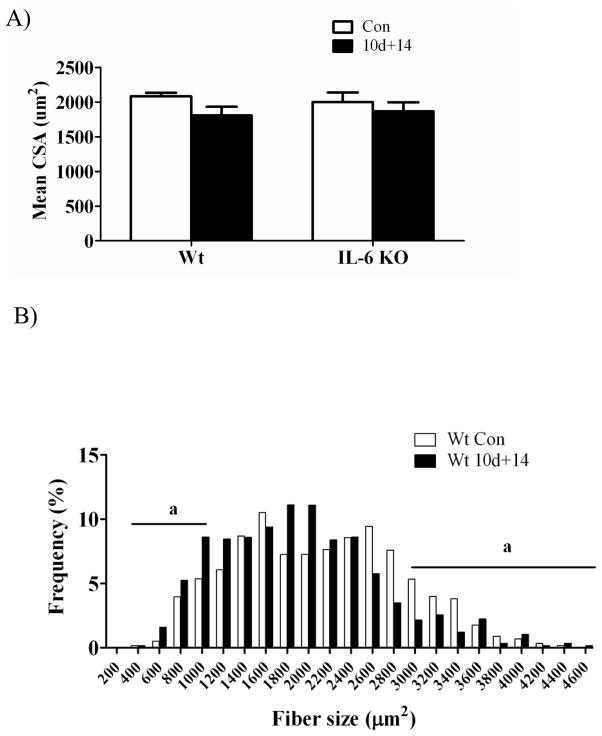
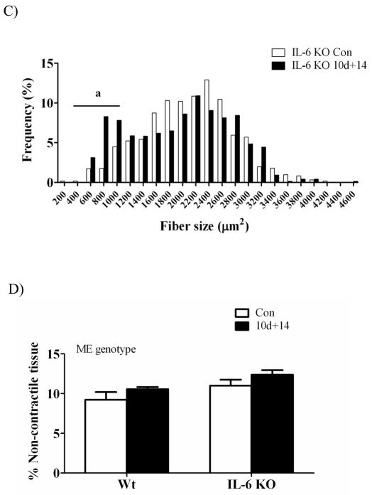
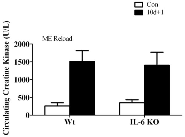
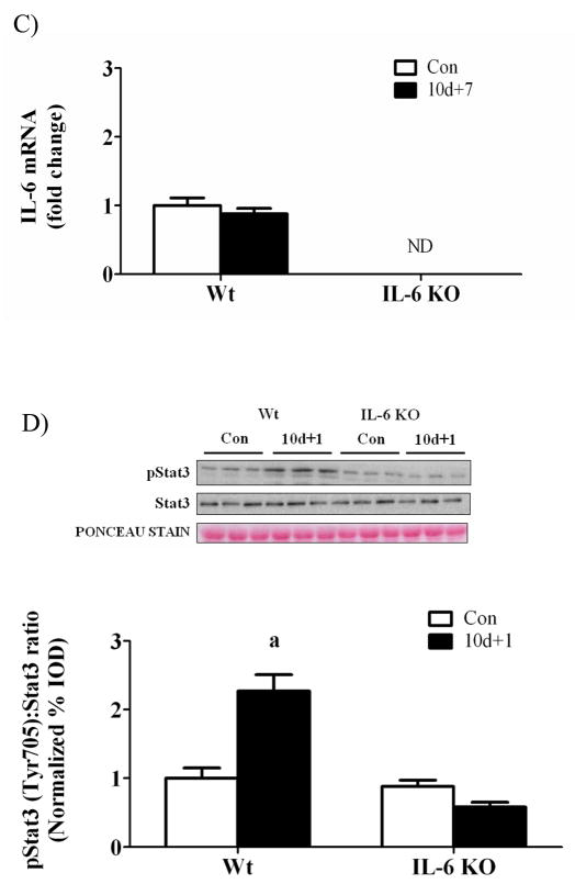
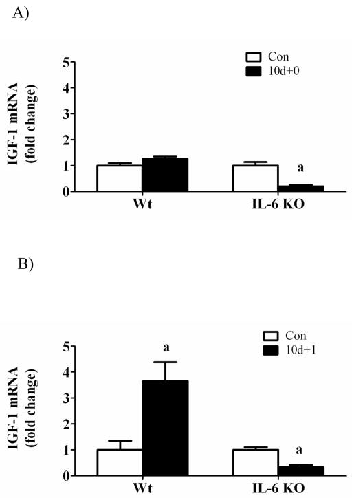
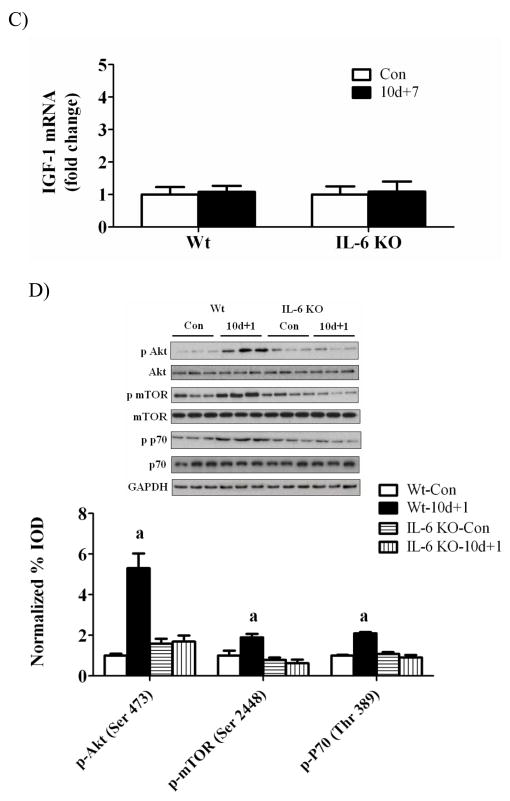
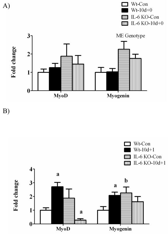
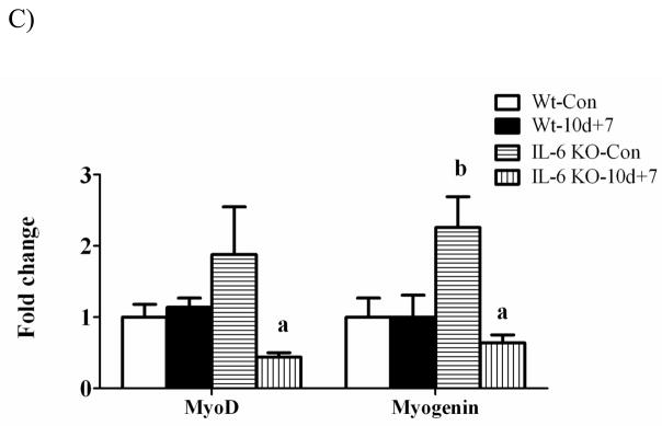
Results: GAS muscle atrophy was induced by 10 days of HS. HS induced a 20% loss of GAS mass in both WT and IL-6 KO mice. HS+7 days of recovery restored WT GAS mass to cage-control values. GAS mass from IL-6 KO mice did not return to cage-control values until HS+14 days of recovery. Both IGF-1 mRNA expression and Akt/mTOR signalling were increased in WT muscle after 1 day of recovery. In IL-6 KO muscle, IGF-1 mRNA expression was decreased and Akt/mTOR signalling was not induced after 1 day of recovery. MyoD and myogenin mRNA expression were both induced in WT muscle after 1 day of recovery, but not in IL-6 KO muscle.

Conclusion: Muscle IL-6 expression appears important for the initial growth response during the recovery from disuse.


Conflict of interest statement

Conflicts of Interest

There is no conflicts of interest.

Figures

Figure 1
Figure 1
Myofiber cross sectional area, fiber distribution, and % non-contractile tissue in gastrocnemius muscle after 2 weeks of recovery from disuse in wild-type mice and IL-6 KO mice. A) Mean myofiber cross sectional area. B) The effect of 14 days of reloading following 10 days of hindlimb suspension on gastrocnemius myofiber size distribution in wild-type mice. C) The effect of 14 days of reloading following 10 days of hindlimb suspension on gastrocnemius myofiber size distribution in IL-6 KO mice. D) The effect of 14 days of reloading following 10 days of hindlimb suspension on % of non-contractile tissue. Values are means ± SE, n= 4–8 per group. a, significant difference in the % of small and/or large fibers. ME, main effect.
Figure 1
Figure 1
Myofiber cross sectional area, fiber distribution, and % non-contractile tissue in gastrocnemius muscle after 2 weeks of recovery from disuse in wild-type mice and IL-6 KO mice. A) Mean myofiber cross sectional area. B) The effect of 14 days of reloading following 10 days of hindlimb suspension on gastrocnemius myofiber size distribution in wild-type mice. C) The effect of 14 days of reloading following 10 days of hindlimb suspension on gastrocnemius myofiber size distribution in IL-6 KO mice. D) The effect of 14 days of reloading following 10 days of hindlimb suspension on % of non-contractile tissue. Values are means ± SE, n= 4–8 per group. a, significant difference in the % of small and/or large fibers. ME, main effect.
Figure 2
Figure 2
The effect of recovery day following disuse-induced atrophy on skeletal muscle damage. The effect of 1 day of reloading following 10 days of disuse-induced atrophy on circulating creatine kinase activity. Circulating creatine kinase activity is expressed as Units per liter (U/L). Values are means ± SE, n = 3–8 per group. ME, main effect.
Figure 3
Figure 3
IL-6 signaling during 0, 1, and 7 days of reloading following 10 days of hindlimb suspension in wild-type and IL-6 KO mice. A) The effect of 10 days of hindlimb suspension on IL-6 mRNA abundance in the gastrocnemius muscle. B) The effect of 1 day of reloading following 10 days of hindlimb suspension on IL-6 mRNA abundance in the gastrocnemius muscle. C) The effect of 7 days of reloading following 10 days of hindlimb suspension on IL-6 mRNA abundance in the gastrocnemius muscle. D). Upper representative western blot of phospho and total forms of Stat3. Lower The ratio of phospho and total Stat3. Values are means ± SE, n = 4–6 per group. a, significantly different from same genotype control. ND, not detectable.
Figure 3
Figure 3
IL-6 signaling during 0, 1, and 7 days of reloading following 10 days of hindlimb suspension in wild-type and IL-6 KO mice. A) The effect of 10 days of hindlimb suspension on IL-6 mRNA abundance in the gastrocnemius muscle. B) The effect of 1 day of reloading following 10 days of hindlimb suspension on IL-6 mRNA abundance in the gastrocnemius muscle. C) The effect of 7 days of reloading following 10 days of hindlimb suspension on IL-6 mRNA abundance in the gastrocnemius muscle. D). Upper representative western blot of phospho and total forms of Stat3. Lower The ratio of phospho and total Stat3. Values are means ± SE, n = 4–6 per group. a, significantly different from same genotype control. ND, not detectable.
Figure 4
Figure 4
IGF-1 signaling during 0, 1, and 7 days of reloading following 10 days of hindlimb suspension in WT and IL-6 KO mice. A) The effect of 10 days of hindlimb suspension on IGF-1 mRNA abundance in the gastrocnemius muscle. B) The effect of 1 day of reloading following 10 days of hindlimb suspension on IGF-1 mRNA abundance in the gastrocnemius muscle. C) The effect of 7 days of reloading following 10 days of hindlimb suspension on IGF-1 mRNA abundance in the gastrocnemius muscle. D). Upper representative western blot of phospho and total forms of Akt, mTOR, and p70. Lower The ratio of phospho and total Akt, mTOR and p70 after 1 day of reloading. Values are means ± SE, n = 4–6 per group. a, significantly different from same genotype control.
Figure 4
Figure 4
IGF-1 signaling during 0, 1, and 7 days of reloading following 10 days of hindlimb suspension in WT and IL-6 KO mice. A) The effect of 10 days of hindlimb suspension on IGF-1 mRNA abundance in the gastrocnemius muscle. B) The effect of 1 day of reloading following 10 days of hindlimb suspension on IGF-1 mRNA abundance in the gastrocnemius muscle. C) The effect of 7 days of reloading following 10 days of hindlimb suspension on IGF-1 mRNA abundance in the gastrocnemius muscle. D). Upper representative western blot of phospho and total forms of Akt, mTOR, and p70. Lower The ratio of phospho and total Akt, mTOR and p70 after 1 day of reloading. Values are means ± SE, n = 4–6 per group. a, significantly different from same genotype control.
Figure 5
Figure 5
The effect of reloading on myogenic regulatory factors in the gastrocnemius muscle of wild-type and IL-6 KO. A) The effect of 10 days of hindlimb suspension on MyoD and myogenin mRNA abundance in the gastrocnemius muscle. B) The effect of 1 day of reloading following 10 days of hindlimb suspension on MyoD and myogenin mRNA abundance in the gastrocnemius muscle. C) The effect of 7 days of reloading following 10 days of hindlimb suspension on MyoD and myogenin mRNA abundance in the gastrocnemius muscle. Values are means ± SE, n = 4–6 per group. a, significantly different from same genotype control. b, significantly different from wild-type control group. ME, main effect.
Figure 5
Figure 5
The effect of reloading on myogenic regulatory factors in the gastrocnemius muscle of wild-type and IL-6 KO. A) The effect of 10 days of hindlimb suspension on MyoD and myogenin mRNA abundance in the gastrocnemius muscle. B) The effect of 1 day of reloading following 10 days of hindlimb suspension on MyoD and myogenin mRNA abundance in the gastrocnemius muscle. C) The effect of 7 days of reloading following 10 days of hindlimb suspension on MyoD and myogenin mRNA abundance in the gastrocnemius muscle. Values are means ± SE, n = 4–6 per group. a, significantly different from same genotype control. b, significantly different from wild-type control group. ME, main effect.
Figure 6
Figure 6
The effect of reloading on cyclin D1 in the gastrocnemius muscle of wild-type and IL-6 KO. A) The effect of 10 days of hindlimb suspension on cyclin D1 mRNA abundance in the gastrocnemius muscle. B) The effect of 1 day of reloading following 10 days of hindlimb suspension on cyclin D1 mRNA abundance in the gastrocnemius muscle. C) The effect of 7 days of reloading following 10 days of hindlimb suspension on cyclin D1 mRNA abundance in the gastrocnemius muscle. D). Upper representative western blot of cyclinD1 protein. Lower Quantified cyclinD1 protein expression after 1 day of reloading. Values are means ± SE, n = 4–6 per group. ME, main effect.
Figure 6
Figure 6
The effect of reloading on cyclin D1 in the gastrocnemius muscle of wild-type and IL-6 KO. A) The effect of 10 days of hindlimb suspension on cyclin D1 mRNA abundance in the gastrocnemius muscle. B) The effect of 1 day of reloading following 10 days of hindlimb suspension on cyclin D1 mRNA abundance in the gastrocnemius muscle. C) The effect of 7 days of reloading following 10 days of hindlimb suspension on cyclin D1 mRNA abundance in the gastrocnemius muscle. D). Upper representative western blot of cyclinD1 protein. Lower Quantified cyclinD1 protein expression after 1 day of reloading. Values are means ± SE, n = 4–6 per group. ME, main effect.
Figure 7
Figure 7
The effect of reloading on extracellular matrix gene expression in the gastrocnemius muscle of Wild-type and IL-6 KO. A) The effect of 10 days of hindlimb suspension on procollagen I, procollagen III, fibronectin mRNA abundance in the gastrocnemius muscle. B) The effect of 1 day of reloading following 10 days of hindlimb suspension on procollagen I, procollagen III, fibronectin mRNA abundance in the gastrocnemius muscle. C) The effect of 7 days of reloading following 10 days of hindlimb suspension on procollagen I, procollagen III, fibronectin mRNA abundance in the gastrocnemius muscle. Values are means ± SE, n = 4–6 per group. a, significantly different from same genotype control. b, significantly different from wild-type matched treatment group.ME, main effect.
Figure 7
Figure 7
The effect of reloading on extracellular matrix gene expression in the gastrocnemius muscle of Wild-type and IL-6 KO. A) The effect of 10 days of hindlimb suspension on procollagen I, procollagen III, fibronectin mRNA abundance in the gastrocnemius muscle. B) The effect of 1 day of reloading following 10 days of hindlimb suspension on procollagen I, procollagen III, fibronectin mRNA abundance in the gastrocnemius muscle. C) The effect of 7 days of reloading following 10 days of hindlimb suspension on procollagen I, procollagen III, fibronectin mRNA abundance in the gastrocnemius muscle. Values are means ± SE, n = 4–6 per group. a, significantly different from same genotype control. b, significantly different from wild-type matched treatment group.ME, main effect.

Similar articles

Cited by

References

    1. Adams GR. Role of insulin-like growth factor-I in the regulation of skeletal muscle adaptation to increased loading. Exerc Sport Sci Rev. 1998;26:31–60. - PubMed
    1. Adams GR. Autocrine and/or paracrine insulin-like growth factor-I activity in skeletal muscle. Clin Orthop Relat Res. 2002:S188–96. - PubMed
    1. Adams GR, Haddad F. The relationships among IGF-1, DNA content, and protein accumulation during skeletal muscle hypertrophy. J Appl Physiol. 1996;81:2509–16. - PubMed
    1. Adams GR, Haddad F, Baldwin KM. Time course of changes in markers of myogenesis in overloaded rat skeletal muscles. J Appl Physiol. 1999;87:1705–12. - PubMed
    1. Ahtikoski AM, Koskinen SO, Virtanen P, Kovanen V, Takala TE. Regulation of synthesis of fibrillar collagens in rat skeletal muscle during immobilization in shortened and lengthened positions. Acta Physiol Scand. 2001;172:131–40. - PubMed

Publication types