Chromosomal variability of human mesenchymal stem cells cultured under hypoxic conditions
- PMID: 21418515
- PMCID: PMC3823094
- DOI: 10.1111/j.1582-4934.2011.01303.x
Chromosomal variability of human mesenchymal stem cells cultured under hypoxic conditions
Abstract
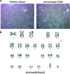
Bone marrow derived human mesenchymal stem cells (hMSCs) have attracted great interest from both bench and clinical researchers because of their pluripotency and ease of expansion ex vivo. However, these cells do finally reach a senescent stage and lose their multipotent potential. Proliferation of these cells is limited up to the time of their senescence, which limits their supply, and they may accumulate chromosomal changes through ex vivo culturing. The safe, rapid expansion of hMSCs is critical for their clinical application. Chromosomal aberration is known as one of the hallmarks of human cancer, and therefore it is important to understand the chromosomal stability and variability of ex vivo expanded hMSCs before they are used widely in clinical applications. In this study, we examined the effects of culturing under ambient (20%) or physiologic (5%) O(2) concentrations on the rate of cell proliferation and on the spontaneous transformation of hMSCs in primary culture and after expansion, because it has been reported that culturing under hypoxic conditions accelerates the propagation of hMSCs. Bone marrow samples were collected from 40 patients involved in clinical research. We found that hypoxic conditions promote cell proliferation more favourably than normoxic conditions. Chromosomal aberrations, including structural instability or aneuploidy, were detected in significantly earlier passages under hypoxic conditions than under normoxic culture conditions, suggesting that amplification of hMSCs in a low-oxygen environment facilitated chromosomal instability. Furthermore, smoothed hazard-function modelling of chromosomal aberrations showed increased hazard after the fourth passage under both sets of culture conditions, and showed a tendency to increase the detection rate of primary karyotypic abnormalities among donors aged 60 years and over. In conclusion, we propose that the continuous monitoring of hMSCs will be required before they are used in therapeutic applications in the clinic, especially when cells are cultured under hypoxic conditions.
© 2011 The Authors Journal of Cellular and Molecular Medicine © 2011 Foundation for Cellular and Molecular Medicine/Blackwell Publishing Ltd.
Figures


Similar articles
-
Donor age and long-term culture affect differentiation and proliferation of human bone marrow mesenchymal stem cells.Ann Hematol. 2012 Aug;91(8):1175-86. doi: 10.1007/s00277-012-1438-x. Epub 2012 Mar 8. Ann Hematol. 2012. PMID: 22395436
-
Chromosomal stability of mesenchymal stromal cells during in vitro culture.Cytotherapy. 2016 Mar;18(3):336-43. doi: 10.1016/j.jcyt.2015.11.017. Epub 2016 Jan 15. Cytotherapy. 2016. PMID: 26780865 Free PMC article.
-
Human bone marrow mesenchymal stem cells support the derivation and propagation of human induced pluripotent stem cells in culture.Cell Reprogram. 2013 Jun;15(3):216-23. doi: 10.1089/cell.2012.0064. Cell Reprogram. 2013. PMID: 23713432 Free PMC article.
-
[Safety evaluation of tissue engineered medical devices using normal human mesenchymal stem cells].Yakugaku Zasshi. 2007 May;127(5):851-6. doi: 10.1248/yakushi.127.851. Yakugaku Zasshi. 2007. PMID: 17473528 Review. Japanese.
-
Scaling up human mesenchymal stem cell manufacturing using bioreactors for clinical uses.Curr Res Transl Med. 2023 Apr-Jun;71(2):103393. doi: 10.1016/j.retram.2023.103393. Epub 2023 Apr 28. Curr Res Transl Med. 2023. PMID: 37163885 Review.
Cited by
-
Differentiation Potential and Tumorigenic Risk of Rat Bone Marrow Stem Cells Are Affected By Long-Term In Vitro Expansion.Turk J Haematol. 2019 Nov 18;36(4):255-265. doi: 10.4274/tjh.galenos.2019.2019.0100. Epub 2019 Jul 9. Turk J Haematol. 2019. PMID: 31284704 Free PMC article.
-
Differentiation of umbilical cord lining membrane-derived mesenchymal stem cells into endothelial-like cells.Iran Biomed J. 2014;18(2):67-75. doi: 10.6091/ibj.1261.2013. Iran Biomed J. 2014. PMID: 24518546 Free PMC article.
-
Origin of Cancer: An Information, Energy, and Matter Disease.Front Cell Dev Biol. 2016 Nov 17;4:121. doi: 10.3389/fcell.2016.00121. eCollection 2016. Front Cell Dev Biol. 2016. PMID: 27909692 Free PMC article.
-
Identification of new genes associated to senescent and tumorigenic phenotypes in mesenchymal stem cells.Sci Rep. 2017 Dec 19;7(1):17837. doi: 10.1038/s41598-017-16224-5. Sci Rep. 2017. PMID: 29259202 Free PMC article.
-
Senescence in Human Mesenchymal Stem Cells: Functional Changes and Implications in Stem Cell-Based Therapy.Int J Mol Sci. 2016 Jul 19;17(7):1164. doi: 10.3390/ijms17071164. Int J Mol Sci. 2016. PMID: 27447618 Free PMC article. Review.
References
-
- Lee OK, Kuo TK, Chen WM, et al. Isolation of multipotent mesenchymal stem cells from umbilical cord blood. Blood. 2004;103:1669–75. - PubMed
-
- Bruder SP, Jaiswal N, Haynesworth SE. Growth kinetics, self-renewal, and the osteogenic potential of purified human mesenchymal stem cells during extensive subcultivation and following cryopreservation. J Cell Biochem. 1997;64:278–94. - PubMed
-
- Jaiswal N, Haynesworth SE, Caplan AI, et al. Osteogenic differentiation of purified, culture-expanded human mesenchymal stem cells in vitro. J Cell Biochem. 1997;64:295–312. - PubMed
-
- Mackay AM, Beck SC, Murphy JM, et al. Chondrogenic differentiation of cultured human mesenchymal stem cells from marrow. Tissue Eng. 1998;4:415–28. - PubMed
Publication types
MeSH terms
Substances
LinkOut - more resources
Full Text Sources

