. 2011 Mar 18:10:29.
doi: 10.1186/1476-4598-10-29.

miR-449 inhibits cell proliferation and is down-regulated in gastric cancer

Affiliations

miR-449 inhibits cell proliferation and is down-regulated in gastric cancer

Tony Bou Kheir et al. Mol Cancer. .

Abstract

Background: Gastric cancer is the fourth most common cancer in the world and the second most prevalent cause of cancer related death. The development of gastric cancer is mainly associated with H. Pylori infection leading to a focus in pathology studies on bacterial and environmental factors, and to a lesser extent on the mechanistic development of the tumour. MicroRNAs are small non-coding RNA molecules involved in post-transcriptional gene regulation. They are found to regulate genes involved in diverse biological functions and alterations in microRNA expression have been linked to the pathogenesis of many malignancies. The current study is focused on identifying microRNAs involved in gastric carcinogenesis and to explore their mechanistic relevance by characterizing their targets.

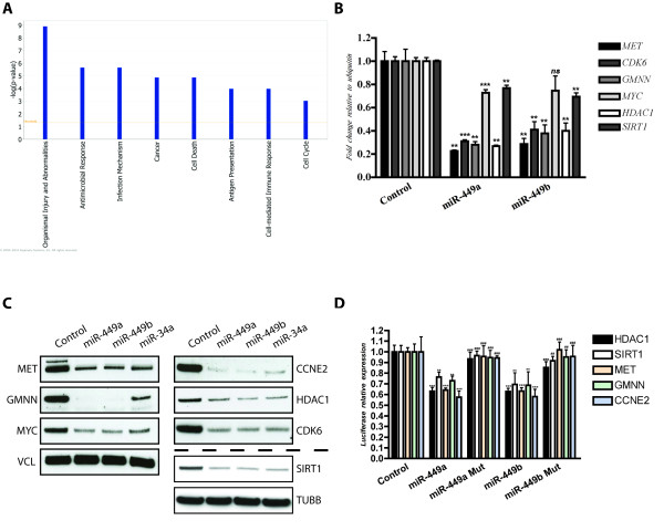
Results: Invitrogen NCode miRNA microarrays identified miR-449 to be decreased in 1-year-old Gastrin KO mice and in H. Pylori infected gastric tissues compared to tissues from wild type animals. Growth rate of gastric cell lines over-expressing miR-449 was inhibited by 60% compared to controls. FACS cell cycle analysis of miR-449 over-expressing cells showed a significant increase in the sub-G1 fraction indicative of apoptosis. ß-Gal assays indicated a senescent phenotype of gastric cell lines over-expressing miR-449. Affymetrix 133v2 arrays identified GMNN, MET, CCNE2, SIRT1 and CDK6 as miR-449 targets. Luciferase assays were used to confirm GMNN, MET, CCNE2 and SIRT1 as direct targets. We also show that miR-449 over-expression activated p53 and its downstream target p21 as well as the apoptosis markers cleaved CASP3 and PARP. Importantly, qPCR analyses showed a loss of miR-449 expression in human clinical gastric tumours compared to normal tissues.

Conclusions: In this study, we document a diminished expression of miR-449 in Gastrin KO mice and further confirmed its loss in human gastric tumours. We investigated the function of miR-449 by identifying its direct targets. Furthermore we show that miR-449 induces senescence and apoptosis by activating the p53 pathway.


Figures

Figure 1
Figure 1
Old Gastrin knockout mice develop gastric adenomas. Antrum sections isolated from 12-16 weeks old mice (left panel), 12 to 18 months old mice (middle panel) and older than 18 months mice (right panel), from wild type (upper panel) and Gastrin knockout mice (lower panel). Sections show adenoma development in antrum tissues in the Gastrin knockouts compared to the wild types.
Figure 2
Figure 2
miR-449 is part of the miR-34 family and inhibits cell proliferation. A - miR-449 is part of the miR-34 family and is evolutionarily conserved. B - miR-449 re-introduction into human gastric cell lines (SNU638) inhibits cell proliferation (red line) compared to a scrambled control (blue line) and miR-146 control (black line). Error bars represent S.D. C - Visual inspection of cell proliferation inhibition and senescence-like phenotype upon miR-449 re-introduction into SNU638 cells (lower panel) compared to scrambled transfection control (upper panel). D - FACS cell cycle analysis showing sub-G1 accumulation of SNU638 cells 72 hours following miR-449 re-introduction (right histogram) compared to a scrambled transfection control (left histogram) The table shows cell accumulation in G1 fraction upon miR-449 re-introduction compared to scrambled RNA control at 48 hours post transfection, followed by a shift to the sub-G1 fraction at 72 hours post transfection indicative of cell death. E - Senescent phenotype of SNU638 cells upon miR-449 and miR-34a positive control re-introduction shown by acidic β-gal assay compared to RNA scrambled control.
Figure 3
Figure 3
miR-449 targets cell cycle controller genes. A - Ingenuity Pathway Analysis (IPA) of deregulated genes upon miR-449 re-introduction into SNU638 cells showing enrichment for the gene categories cancer, cell death and cell cycle pathways among others. B - qPCR validation of Affymetrix arrays showing down-regulation of MET, CDK6, GMNN, MYC and HDAC1 upon miR-449 re-introduction compared to scrambled RNA controls. C - Western blot validation of down-regulated genes upon miR-449 re-introduction into SNU638 cells compared to scrambled RNA controls. Vinculin (VCL) and tubulin beta (TUBB) were used as loading controls D - verification of direct and functional target binding using luciferase constructs holding wild type 3'UTRs and mutated 3'UTRs (two mutations in miR-449 binding site), * indicates statistical significance in luciferase expression between wild type 3'UTRs transfected with miR449a/b compared to RNA scrambled control, # indicates statistical difference in luciferase expression between wild type 3'UTRs compared to mutant 3'UTRs transfected with miR-449a and miR-449b. "ns" not significant p value > 0.05, "*" or "#" significant 0.01 < p value < 0.05, "**" or "##" very significant 0.01 < p value < 0.001, "***" or "###" extremely significant p value < 0.001
Figure 4
Figure 4
miR-449 activates the p53 pathway. Western blot analysis showing an increase of the p53 protein upon miR-449 and positive control miR-34a re-introduction into SNU638 cells compared to RNA scrambled control as well as an activation of the p53 downstream target p21 and apoptosis markers cleaved CASP3 and PARP. Vinculin (VCL) and tubulin beta (TUBB) were used as loading controls.
Figure 5
Figure 5
miR-449 is down regulated in human gastric cancers. qPCR analysis (upper panel) showing down-regulation of miR-449 expression in 8 gastric cancer tissues compared to miR-449 expression in sample-matched controls (dotted line). U44 was used as endogenous control. Table showing clinical information of patients (lower panel). "ns" not significant p value > 0.05, "*" significant 0.01 < p value < 0.05, "**" very significant 0.01 < p value < 0.001, "***" extremely significant p value < 0.001

Similar articles

Cited by

References

    1. Smith MG, Hold GL, Tahara E, El-Omar EM. Cellular and molecular aspects of gastric cancer. World J Gastroenterol. 2006;12:2979–2990. - PMC - PubMed
    1. Kountouras J, Zavos C, Chatzopoulos D, Katsinelos P. New aspects of Helicobacter pylori infection involvement in gastric oncogenesis. J Surg Res. 2008;146:149–158. doi: 10.1016/j.jss.2007.06.011. - DOI - PubMed
    1. Polk DB, Peek RM Jr. Helicobacter pylori: gastric cancer and beyond. Nat Rev Cancer. 2010;10:403–414. doi: 10.1038/nrc2857. - DOI - PMC - PubMed
    1. Wang TC, Dangler CA, Chen D, Goldenring JR, Koh T, Raychowdhury R, Coffey RJ, Ito S, Varro A, Dockray GJ, Fox JG. Synergistic interaction between hypergastrinemia and Helicobacter infection in a mouse model of gastric cancer. Gastroenterology. 2000;118:36–47. doi: 10.1016/S0016-5085(00)70412-4. - DOI - PubMed
    1. Takaishi S, Tu S, Dubeykovskaya ZA, Whary MT, Muthupalani S, Rickman BH, Rogers AB, Lertkowit N, Varro A, Fox JG, Wang TC. Gastrin is an essential cofactor for helicobacter-associated gastric corpus carcinogenesis in C57BL/6 mice. Am J Pathol. 2009;175:365–375. doi: 10.2353/ajpath.2009.081165. - DOI - PMC - PubMed

Publication types

MeSH terms