

Biological and immunological characterization of recombinant Yellow Fever 17D viruses expressing a Trypanosoma cruzi Amastigote Surface Protein-2 CD8+ T cell epitope at two distinct regions of the genome
- PMID: 21418577
- PMCID: PMC3066119
- DOI: 10.1186/1743-422X-8-127
Biological and immunological characterization of recombinant Yellow Fever 17D viruses expressing a Trypanosoma cruzi Amastigote Surface Protein-2 CD8+ T cell epitope at two distinct regions of the genome
Abstract
Background: The attenuated Yellow fever (YF) 17D vaccine virus is one of the safest and most effective viral vaccines administered to humans, in which it elicits a polyvalent immune response. Herein, we used the YF 17D backbone to express a Trypanosoma cruzi CD8+ T cell epitope from the Amastigote Surface Protein 2 (ASP-2) to provide further evidence for the potential of this virus to express foreign epitopes. The TEWETGQI CD8+ T cell epitope was cloned and expressed based on two different genomic insertion sites: in the fg loop of the viral Envelope protein and the protease cleavage site between the NS2B and NS3. We investigated whether the site of expression had any influence on immunogenicity of this model epitope.
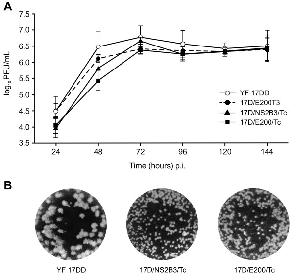
Results: Recombinant viruses replicated similarly to vaccine virus YF 17D in cell culture and remained genetically stable after several serial passages in Vero cells. Immunogenicity studies revealed that both recombinant viruses elicited neutralizing antibodies to the YF virus as well as generated an antigen-specific gamma interferon mediated T-cell response in immunized mice. The recombinant viruses displayed a more attenuated phenotype than the YF 17DD vaccine counterpart in mice. Vaccination of a mouse lineage highly susceptible to infection by T. cruzi with a homologous prime-boost regimen of recombinant YF viruses elicited TEWETGQI specific CD8+ T cells which might be correlated with a delay in mouse mortality after a challenge with a lethal dose of T. cruzi.
Conclusions: We conclude that the YF 17D platform is useful to express T. cruzi (Protozoan) antigens at different functional regions of its genome with minimal reduction of vector fitness. In addition, the model T. cruzi epitope expressed at different regions of the YF 17D genome elicited a similar T cell-based immune response, suggesting that both expression sites are useful. However, the epitope as such is not protective and it remains to be seen whether expression of larger domains of ASP-2, which include the TEWETGQI epitope, will elicit better T-CD8+ responses to the latter. It is likely that additional antigens and recombinant virus formulations will be necessary to generate a protective response.
Figures


Similar articles
-
Vaccination using recombinants influenza and adenoviruses encoding amastigote surface protein-2 are highly effective on protection against Trypanosoma cruzi infection.PLoS One. 2013 Apr 24;8(4):e61795. doi: 10.1371/journal.pone.0061795. Print 2013. PLoS One. 2013. PMID: 23637908 Free PMC article.
-
Recombinant yellow fever viruses elicit CD8+ T cell responses and protective immunity against Trypanosoma cruzi.PLoS One. 2013;8(3):e59347. doi: 10.1371/journal.pone.0059347. Epub 2013 Mar 19. PLoS One. 2013. PMID: 23527169 Free PMC article.
-
Immune responses against a single CD8+-T-cell epitope induced by virus vector vaccination can successfully control Trypanosoma cruzi infection.Infect Immun. 2005 Nov;73(11):7356-65. doi: 10.1128/IAI.73.11.7356-7365.2005. Infect Immun. 2005. PMID: 16239534 Free PMC article.
-
Live virus vaccines based on a yellow fever vaccine backbone: standardized template with key considerations for a risk/benefit assessment.Vaccine. 2015 Jan 1;33(1):62-72. doi: 10.1016/j.vaccine.2014.10.004. Epub 2014 Oct 27. Vaccine. 2015. PMID: 25446819 Free PMC article. Review.
-
The yellow fever 17D virus as a platform for new live attenuated vaccines.Hum Vaccin Immunother. 2014;10(5):1256-65. doi: 10.4161/hv.28117. Epub 2014 Feb 19. Hum Vaccin Immunother. 2014. PMID: 24553128 Free PMC article. Review.
Cited by
-
Advances and challenges towards a vaccine against Chagas disease.Hum Vaccin. 2011 Nov;7(11):1184-91. doi: 10.4161/hv.7.11.17016. Epub 2011 Nov 1. Hum Vaccin. 2011. PMID: 22048121 Free PMC article. Review.
-
A novel therapeutic HBV vaccine candidate induces strong polyfunctional cytotoxic T cell responses in mice.JHEP Rep. 2021 Apr 22;3(4):100295. doi: 10.1016/j.jhepr.2021.100295. eCollection 2021 Aug. JHEP Rep. 2021. PMID: 34159304 Free PMC article.
-
Vaccination using recombinants influenza and adenoviruses encoding amastigote surface protein-2 are highly effective on protection against Trypanosoma cruzi infection.PLoS One. 2013 Apr 24;8(4):e61795. doi: 10.1371/journal.pone.0061795. Print 2013. PLoS One. 2013. PMID: 23637908 Free PMC article.
-
Yellow Fever 17DD Vaccine Virus Infection Causes Detectable Changes in Chicken Embryos.PLoS Negl Trop Dis. 2015 Sep 15;9(9):e0004064. doi: 10.1371/journal.pntd.0004064. eCollection 2015. PLoS Negl Trop Dis. 2015. PMID: 26371874 Free PMC article.
-
A Yellow Fever 17D Virus Replicon-Based Vaccine Platform for Emerging Coronaviruses.Vaccines (Basel). 2021 Dec 16;9(12):1492. doi: 10.3390/vaccines9121492. Vaccines (Basel). 2021. PMID: 34960238 Free PMC article.
References
-
- Lindenbach B, Thiel HJ, Rice CM. In: Fields Virology. 5. Knipe DM HP, Griffin DE, Martin MA, Lamb RA, Roizman B, Straus SE, editor. Philadelphia: Wolters Kluwer, Lippincott Williams and Wilkins; 2007. Flaviviridae: the viruses and their replication; pp. 1101–1152.
-
- Monath T. In: Vaccines. 4. Plotkin SAO W, editor. Philadelphia: WB Saunders; 2004. Yellow fever vaccine; pp. 1095–1176.
-
- Reinhardt B, Jaspert R, Niedrig M, Kostner C, L'age-Stehr J. Development of viremia and humoral and cellular parameters of immune activation after vaccination with yellow fever virus strain 17D: a model of human flavivirus infection. J Med Virol. 1998;56:159–167. doi: 10.1002/(SICI)1096-9071(199810)56:2<159::AID-JMV10>3.0.CO;2-B. - DOI - PubMed
-
- Co M, Terajima M, Cruz J, Ennis F, Rothman A. Human cytotoxic T lymphocyte responses to live attenuated 17D yellow fever vaccine: identification of HLA-B35-restricted CTL epitopes on nonstructural proteins NS1, NS2b, NS3, and the structural protein E. Virology. 2002;293:151–163. doi: 10.1006/viro.2001.1255. - DOI - PubMed
Publication types
MeSH terms
Substances
LinkOut - more resources
Full Text Sources
Other Literature Sources
Medical
Research Materials
