

Vomiting and wasting disease associated with hemagglutinating encephalomyelitis viruses infection in piglets in Jilin, China
- PMID: 21418610
- PMCID: PMC3071789
- DOI: 10.1186/1743-422X-8-130
Vomiting and wasting disease associated with hemagglutinating encephalomyelitis viruses infection in piglets in Jilin, China
Abstract
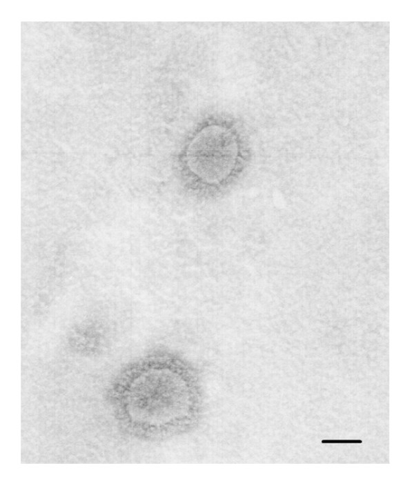
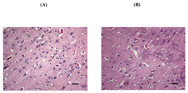
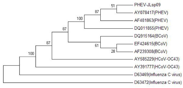
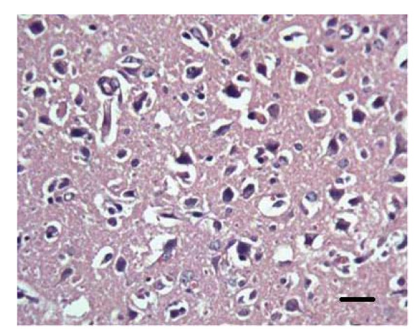
One coronavirus strain was isolated from brain tissues of ten piglets with evident clinical manifestations of vomiting, diarrhea and dyskinesia in Jilin province in China. Antigenic and genomic characterizations of the virus (isolate PHEV-JLsp09) were based on multiplex PCR and negative staining electron microscopy and sequence analysis of the Hemagglutinin-esterase (HE) gene. These piglets were diagnosed with Porcine hemagglutinating encephalomyelitis virus (PHEV).Necropsy was performed on the piglets. Major pathological changes included meningeal hyperemia, meningeal hemorrhage and cortical hemorrhage. Minor changes were also observed in other organs. Histopathological changes included satellitosis and neuronophagia in the cerebral cortex.Mice were infected with the isolated virus. Their histopathological changes were similar to those symptoms observed in the piglets, exhibiting typical changes for non-suppurative encephalitis. Thus, Porcine hemagglutinating encephalomyelitis virus mainly causes damage to the nervous system but also impacts other organs. This viral strain (isolate PHEV-JLsp09) found in the Siping area of Jilin Province in China is evolutionally closest to the HEV-67N stain (North American strain), indicating that this viral strain evolved from the PHEV from North America.
Figures

References
-
- Werdin RE, Sorensen DK, Stewart WC. Porcine encephalomyelitis caused by hemagglutinating encephalomyelitis virus. J Am Vet Med Assoc. 1976;168:240–6. - PubMed
-
- Mengeling WL. Incidence of antibody for hemagglutinating encephalomyelitis virus in serums from swine in the United States. Am J Vet Res. 1975;36:821–823. - PubMed
-
- Straw BL, Zimmerman JJ, Allaire DS, Taylor DJ, editors. Diseases of swine. 9. Ames (IA): Blackwell Publishers; 2006. Pensaert MB Hemagglutinating encephalomyelitis virus; pp. 353–358.
Publication types
MeSH terms
LinkOut - more resources
Full Text Sources
Medical
