. 2011 May 15;353(2):331-43.
doi: 10.1016/j.ydbio.2011.03.008. Epub 2011 Mar 16.

Nav2 hypomorphic mutant mice are ataxic and exhibit abnormalities in cerebellar development

Affiliations

Nav2 hypomorphic mutant mice are ataxic and exhibit abnormalities in cerebellar development

Elizabeth M McNeill et al. Dev Biol. .

Abstract

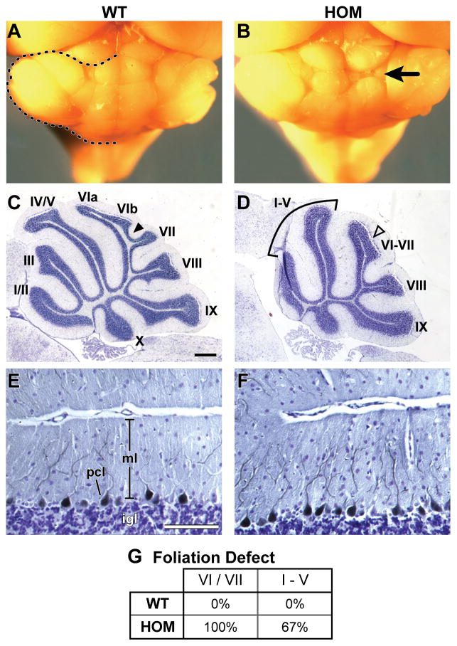
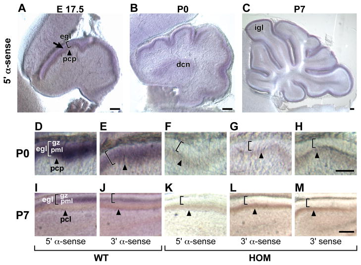
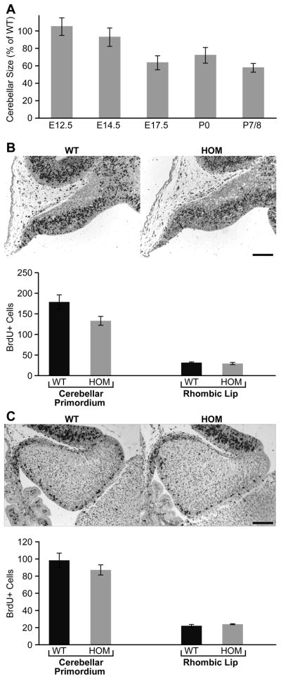
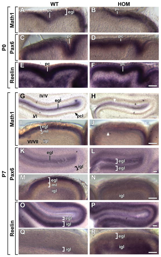
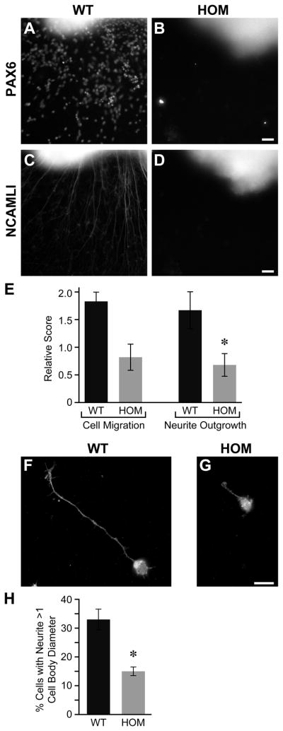
Development of the cerebellum involves a coordinated program of neuronal process outgrowth and migration resulting in a foliated structure that plays a key role in motor function. Neuron navigator 2 (Nav2) is a cytoskeletal-interacting protein that functions in neurite outgrowth and axonal elongation. Herein we show that hypomorphic mutant mice lacking the full-length Nav2 transcript exhibit ataxia and defects in cerebellar development. At embryonic day (E)17.5, the mutant cerebellum is reduced in size and exhibits defects in vermal foliation. Reduction in cell proliferation at early times (E12.5 and E14.5) may contribute to this size reduction. The full-length Nav2 transcript is expressed in the premigratory zone of the external granule layer (EGL). Granule cells in the germinal zone of the EGL appear to proliferate normally, however, due to the reduction in cerebellar circumference there are fewer total BrdU-labeled granule cells in the mutants, and these fail to migrate normally toward the interior of the cerebellum. In Nav2 hypomorphs, fewer granule cells migrate out of cerebellar EGL explants and neurite outgrowth from both explants and isolated external granule cell cultures is reduced. This suggests that the formation of parallel axon fibers and neuronal migration is disrupted in Nav2 mutants. This work supports an essential role for full-length Nav2 in cerebellar development, including axonal elongation and migration of the EGL neurons.


Figures

Figure 1
Figure 1
Nav2−/− hypomorphic mutants exhibit deficits in motor coordination and balance in behavioral tests. (A–C) Gait analysis: mutant mice are off center, take an increased total number of steps and display an increased side width on the left side. (D–E) Mutants display an increased distance traveled on the balance beam and an increased number of missteps relative to wild-type. (F–H) Constant and accelerating rotorod test: mutant mice have a decreased latency to fall and the speed at time of drop is decreased from wild-type animals. (I–K) Nav2−/− hypomorphic mutants exhibit deficits in coordination and activity by visual observation in stereotyped behavioral analysis including a decreased percentage of time rearing, and increased percentage of time rotating as well as an increased percentage of time active. * Indicates a p value ≤ 0.05; ** indicates a p value ≤ 0.01; values are mean ± standard error of the mean.
Figure 2
Figure 2
Cerbellar abnormalities in adult hypomorphic Nav2−/− mutants. (A) Wild-type cerebellum (dotted line outlines the left side of the cerebellum). B) Abnormal vermal folding on the right side of an adult Nav2 mutant cerebellum (black arrow). (C) Normal foliation in the wild-type animal in (A) is shown in a medial cresyl violet-stained sagittal section (10 μm). Note the presence of the intercural fissue (closed arrowhead) separating folia VIb and VII. (D) Abnormal foliation in the mutant shown in (B); note the presence of only 2 folia in the region of I–V in the homozygote compared to 3 distinct folia (I/II, III, and IV/V) in the wild-type; the homozygote also shows a reduction in overall development of VI/VII including the absence of the intercural fissure (open arrowhead, D). (E,F) Overall cytoarchitecture as revealed by cresyl violet staining. The development of the molecular, Purkinje (stained with antibody to calbindin d28k), and granule layers (dark blue). Note the closer spacing of the Purkinje cell bodies in the mutant. (G) The percentage of animals with a defect in foliation in the regions I–V and VI/VII as determined from analysis of medial sagittal sections is shown for wild-type (n = 6) and homozygous mutant (n = 12) mice. igl, inner granule layer; ml, molecular layer; pcl, Purkinje cell layer. Folia are designated by roman numerals (I/II–X). Scale bars: 100 μm C–F.
Figure 3
Figure 3
Nav2 mRNA expression in the embryonic and postnatal cerebellum. (A–C) The Nav2 transcript in wild-type mice is detected using the 5′ probe in the premigratory granule cells within the EGL (bracket) at E17.5, P0 and P7. Specific staining is also seen in the deep cerebellar nucleus (P0) and the inner granule layer (P7). The location of the Purkinje cell plate (brown) is indicated by the closed arrowhead. Specific Nav2 staining is shown at P0 and P7 in the inner or pre-migratory cell layer (pml) of the wild-type EGL using both the 5′ (D,I) and 3′ probes (E,J). No specific Nav2 stain is observed with the 5′ probe in the homozygote at P0 (F) or P7 (K). No Nav2 staining is detected with the 3′ probe in the homozygote at these times (G,L), nor is any specific staining detected using the 3′ sense control probe (H,M). dcn, deep cerebellar nucleus; igl, inner granule layer, egl, external granule cell layer (bracket); gz, germinal zone; pcl, Purkinje cell layer; pcp, Purkinje cell plate; pml pre-migratory layer. Scale bars: 100 μm.
Figure 4
Figure 4
Morphology of the cerebellum and folia in wild-type and homozygous Nav2 mice through development. (A–H) Sagittal sections (10 μm) through the medial region of the cerebellum stained with cresyl violet. (A,C) Four fissures are seen in the wild-type cerebellum at E17.5 and P0. (B,D) At E17.5 and P0, the homozygote mutant cerebellum is decreased in size, and fissure formation is delayed; at P0, both the preculminate (pc) and primary (pr) fissures are less well developed compared to the wild-type litter mate control (compare B to A, and D to C). (E–H) At P7 and P17, a lack of tertiary fissure initiation (intercural fissue) delineating folia VI from VII is apparent (compare F to E and H to G). Additionally, formation of secondary fissures delineating I/II, III and IV/V is also lacking in the Nav2 mutant. The number of wild-type and homozygous embryos/animals examined at E17.5, P0, P7 and P14 is 7,10,3,3 and 9,11,3 and 4, respectively. cl, central lobe; sec, secondary; pl, posterolateral. Scale bars: 100 μm A–D; 200 μm E–H.
Figure 5
Figure 5
Development of the cerebellar primordium. (A) Cerebellum size decreases in Nav2 mutants relative to their wild-type counterparts with advancing development. (B) BrdU positive cells in the cerebellar primordium and rhombic lip at E12.5 in a wild-type and Nav2 mutant, with the number of labeled cells in each region shown as group averages of wild-type (n=3) and homozygous mutant (n=3) embryos. (C) BrdU positive cells in the cerebellar primordium and rhombic lip at E14.5 in a wild-type and Nav2 mutant, and quantification of BrdU-labeled cells in each of these areas for wild-type (n=3) and homozygous mutant (n=3) embryos. Scale bars: 100 μm B, C.
Figure 6
Figure 6
Purkinje and granule cell abnormalities in Nav2 mutants at P7 and P8. (A) Calbindin staining of Purkinje cells, and magnification of the region (red box) of the intercural fissure at P7 in a wild-type mouse. (B, C) Calbindin staining at P7 and P8 in Nav2 hypomorphic mutants; magnification (red box) of abnormal Purkinje cell and granule cell distribution in the region where folia VI should be branching from VII (open white arrow). (D) Total Purkinje cell number as well as tissue circumference was evaluated in sections (3–9/animal) and averaged for each animal (n=3 WT and 4 HOM), and the number of Purkinje cells per mm is shown. Scale bars: 100 μm A–C.
Figure 7
Figure 7
Migration of the EGL cells is delayed in Nav2−/− hypomorphic mutant mice. (A) Cresyl violet-stained sections showing only a small number of cells remaining in the EGL of wild-type controls at P15–16, and absence of the EGL by P17 (n=3, 2 and 3, respectively). In the homozygous mutant, the EGL is thicker than that of the wild-type group at P15–P16 and persists at P17 (black arrow) (n = 2,1 and 3, respectively). (B) Analysis of labeled granule cells 2 h after administration of BrdU to wild-type (n=3) and homozygous mutant (n=3) pups at P0. BrdU positive cells in the proliferating layer (germinal zone) of the EGL in vermal sections were counted, and the circumference of the cerebellum was measured. The graph (bottom left) shows the number of BrdU-stained cells per 0.5 mm of tissue in homozygous mutants (expressed as a percentage of wild-type) does not differ from the wild-type controls; however, the total number of BrdU-labeled cells in the germinal zone of the entire cerebellum is significantly reduced in Nav2−/− mutants. (C) Granule cell migration is defective in Nav2−/− mutants. The number of BrdU-labeled granule cells remaining in the proliferating layer of the EGL 40–48 h after maternal injection of BrdU were counted at P0 in wild-type (n=3) and Nav2 mutants (n=3). In the wild-type animal (C, left panel) BrdU-labeled granule cells are dispersed throughout the EGL, and also appear to have moved in the direction of the Purkinje cell plate/IGL. In the Nav2 hypomorphic mutant littermate (C, right panel), BrdU-labeled cells remain tightly packed in the outer layer of the EGL. The preculminate fissure is indicated by the open arrow. The graph (bottom right) shows the number of BrdU-labeled cells (expressed per 0.5 mm tissue length) remaining in the proliferating layer of the EGL is significantly greater in Nav2−/− mutants when compared to wild-type controls. * indicates a p value ≤ 0.02. Scale bars: 50 μm A, B; 100 μm C.
Figure 8
Figure 8
In situ hybridization of Math1, Pax6 and Reelin. (A–F) At P0, wild-type and mutant animals exhibit similar staining in both intensity and distribution (n=3 WT and 2–3 HOM; staining in the region of the preculminate (pc) fissure is shown). (G–R) Transcript expression at P7. (G–J) In the mutant, Math1 expression is more dispersed (white arrowheads) indicating a lack of organization in both folia IV/V (compare H to G) and VI–VII (compare J to I) in the mutant. (K–R) A failure to form sharp boundaries of Pax6 and Reelin expression in the mutant EGL and IGL is shown in folia IV/V and VI–VII (n= 3 WT and 3 HOM per probe). egl, external granule layer; igl, inner granule layer; ml, molecular layer; pc, preculminate fissure; Scale bars: 50 μm A–R.
Figure 9
Figure 9
Granule cell axons are less well developed and granule cells do not migrate normally in Nav2 mutants. Granule cells stained with an antibody to Pax6 in wild-type (A) and Nav2 mutant (B) explants after 72 h. Parallel axon fibers stained with NCAM L1 antibody in wild-type (C) and Nav2 mutant (D) explants after 72 h. Note the absence of neurites and migrating cell bodies in this mutant culture. (E) The migration of cells away from the explant and overall neurite outgrowth was scored in cerebellar explants from 3 wild-type and 11 Nav2 mutants and there was a significant reduction in neurite outgrowth in homozygous mutant mice. * Indicates a p value ≤ 0.05; Scale bars: 50 μm A–D. (F–G) Dissociated granule cells from wild-type (n = 3) and mutant (n = 3) animals stained with and antibody to Abi-1. (H) Wild-type mice showed a significantly greater percentage of cells with neurites longer than 1 cell body in diameter. * Indicates a p value ≤ 0.01; Scale bars: 10 μm F–G.

Similar articles

Cited by

References

    1. Akhmanova A, Steinmetz MO. Tracking the ends: a dynamic protein network controls the fate of microtubule tips. Nat Rev. 2008;9:309–322. - PubMed
    1. Bancroft JD, Stevens A. Theory and practice of histological techniques. 3. Churchill Livingstone; London: 1990.
    1. Bompard G, Caron E. Regulation of WASP/WAVE proteins: making a long story short. J Cell Bio. 2004;166:957–962. - PMC - PubMed
    1. Cajal SR. Texture of the nervous system of man and the vertebrates. Springer-Verlag/Wien; New York: 1911. 2000.
    1. Chédotal A. Should I stay or should I go? Becoming a granule cell. Trends Neurosci. 2010;33:163–172. - PubMed

Publication types

MeSH terms