

Elevated levels of NO are localized to distal airways in asthma
- PMID: 21419218
- PMCID: PMC3124865
- DOI: 10.1016/j.freeradbiomed.2011.03.015
Elevated levels of NO are localized to distal airways in asthma
Erratum in
- Free Radic Biol Med. 2013 May;58:45
Abstract
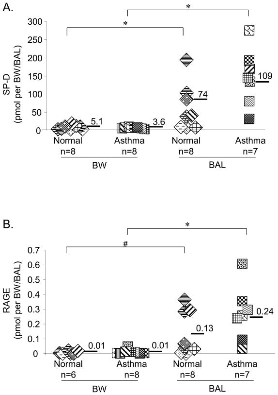
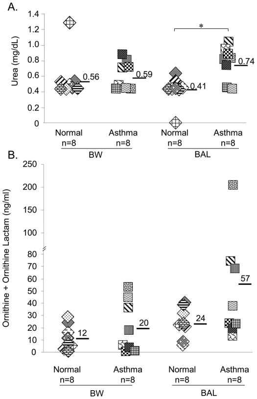
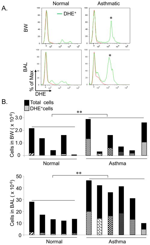
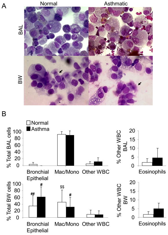
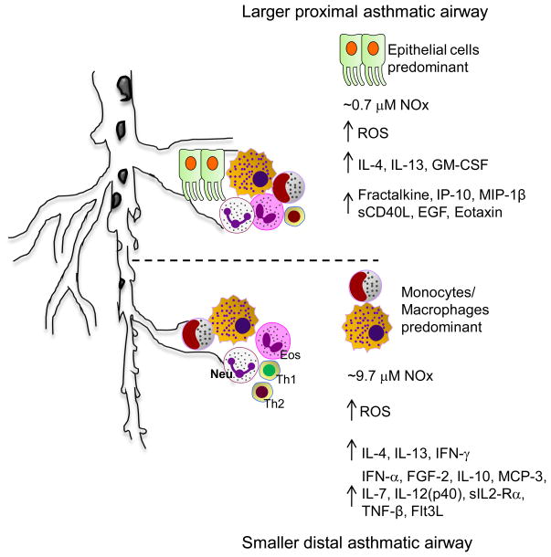
The contribution of nitric oxide (NO) to the pathophysiology of asthma remains incompletely defined despite its established pro- and anti-inflammatory effects. Induction of the inducible nitric oxide synthase (iNOS), arginase, and superoxide pathways is correlated with increased airway hyperresponsiveness in asthmatic subjects. To determine the contributions of these pathways in proximal and distal airways, we compared bronchial wash (BW) to traditional bronchoalveolar lavage (BAL) for measurements of reactive nitrogen/oxygen species, arginase activation, and cytokine/chemokine levels in asthmatic and normal subjects. Levels of NO were preferentially elevated in the BAL, demonstrating higher level NOS activation in the distal airway compartment of asthmatic subjects. In contrast, DHE(+) cells, which have the potential to generate reactive oxygen species, were increased in both proximal and distal airway compartments of asthmatics compared to controls. Different patterns of cytokines and chemokines were observed, with a predominance of epithelial cell-associated mediators in the BW compared to macrophage/monocyte-derived mediators in the BAL of asthmatic subjects. Our study demonstrates differential production of reactive species and soluble mediators within the distal airways compared to the proximal airways in asthma. These results indicate that cellular mechanisms are activated in the distal airways of asthmatics and must be considered in the development of therapeutic strategies for this chronic inflammatory disorder.
Copyright © 2011 Elsevier Inc. All rights reserved.
Figures

Similar articles
-
Inducible nitric oxide synthase expression is increased in the alveolar compartment of asthmatic patients.Allergy. 2017 Apr;72(4):627-635. doi: 10.1111/all.13052. Epub 2016 Oct 14. Allergy. 2017. PMID: 27647044
-
Influence of cigarette smoke on the arginine pathway in asthmatic airways: increased expression of arginase I.J Allergy Clin Immunol. 2007 Feb;119(2):391-7. doi: 10.1016/j.jaci.2006.10.030. J Allergy Clin Immunol. 2007. PMID: 17291856
-
Increased formation of the potent oxidant peroxynitrite in the airways of asthmatic patients is associated with induction of nitric oxide synthase: effect of inhaled glucocorticoid.FASEB J. 1998 Aug;12(11):929-37. FASEB J. 1998. PMID: 9707165 Clinical Trial.
-
[Arginine metabolism in bronchial asthma].Postepy Hig Med Dosw (Online). 2007;61:156-66. Postepy Hig Med Dosw (Online). 2007. PMID: 17410056 Review. Polish.
-
How to measure airway inflammation: bronchoalveolar lavage and airway biopsies.Can Respir J. 1998 Jul-Aug;5 Suppl A:18A-21A. Can Respir J. 1998. PMID: 9753511 Review.
Cited by
-
Myeloid-derived suppressor cells (MDSCs) and mechanistic target of rapamycin (mTOR) signaling pathway interact through inducible nitric oxide synthase (iNOS) and nitric oxide (NO) in asthma.Am J Transl Res. 2019 Sep 15;11(9):6170-6184. eCollection 2019. Am J Transl Res. 2019. PMID: 31632585 Free PMC article.
-
Deficiency of Socs3 leads to brain-targeted EAE via enhanced neutrophil activation and ROS production.JCI Insight. 2019 Apr 2;5(9):e126520. doi: 10.1172/jci.insight.126520. JCI Insight. 2019. PMID: 30939124 Free PMC article.
-
Elevated exhaled nitric oxide in allergen-provoked asthma is associated with airway epithelial iNOS.PLoS One. 2014 Feb 28;9(2):e90018. doi: 10.1371/journal.pone.0090018. eCollection 2014. PLoS One. 2014. PMID: 24587191 Free PMC article.
-
Myeloid-derived suppressor cells function as novel osteoclast progenitors enhancing bone loss in breast cancer.Cancer Res. 2013 Jan 15;73(2):672-82. doi: 10.1158/0008-5472.CAN-12-2202. Epub 2012 Dec 14. Cancer Res. 2013. PMID: 23243021 Free PMC article.
-
Subsets of airway myeloid-derived regulatory cells distinguish mild asthma from chronic obstructive pulmonary disease.J Allergy Clin Immunol. 2015 Feb;135(2):413-424.e15. doi: 10.1016/j.jaci.2014.08.040. Epub 2014 Oct 25. J Allergy Clin Immunol. 2015. PMID: 25420684 Free PMC article.
References
-
- Gelfand EW, Kraft M. The importance and features of the distal airways in children and adults. J Allergy Clin Immunol. 2009;124:S84–87. - PubMed
-
- Grootendorst DC, Sont JK, Willems LN, Kluin-Nelemans JC, Van Krieken JH, Veselic-Charvat M, Sterk PJ. Comparison of inflammatory cell counts in asthma: induced sputum vs bronchoalveolar lavage and bronchial biopsies. Clin Exp Allergy. 1997;27:769–779. - PubMed
-
- Leff A. Pathogenesis of asthma. Neurophysiology and pharmacology of bronchospasm. Chest. 1982;81:224–229. - PubMed
-
- Hyde DM, Hamid Q, Irvin CG. Anatomy, pathology, and physiology of the tracheobronchial tree: emphasis on the distal airways. J Allergy Clin Immunol. 2009;124:S72–77. - PubMed
-
- Dweik RA, Sorkness RL, Wenzel S, Hammel J, Curran-Everett D, Comhair SA, Bleecker E, Busse W, Calhoun WJ, Castro M, Chung KF, Israel E, Jarjour N, Moore W, Peters S, Teague G, Gaston B, Erzurum SC. Use of exhaled nitric oxide measurement to identify a reactive, at-risk phenotype among patients with asthma. Am J Respir Crit Care Med. 181:1033–1041. - PMC - PubMed
Publication types
MeSH terms
Substances
Grants and funding
- S10 RR019231/RR/NCRR NIH HHS/United States
- T32 AI007051/AI/NIAID NIH HHS/United States
- R01 CA131653/CA/NCI NIH HHS/United States
- P30 AT050948/AT/NCCIH NIH HHS/United States
- 5UL1RR025777/RR/NCRR NIH HHS/United States
- P01 HL073907/HL/NHLBI NIH HHS/United States
- UL1 RR025777/RR/NCRR NIH HHS/United States
- P30 DK079337/DK/NIDDK NIH HHS/United States
- HL073907/HL/NHLBI NIH HHS/United States
- F32 HL095341/HL/NHLBI NIH HHS/United States
- S10 RR19231/RR/NCRR NIH HHS/United States
- 1F32HL095341-01A1/HL/NHLBI NIH HHS/United States
- CA131653/CA/NCI NIH HHS/United States
- U54 CA100949/CA/NCI NIH HHS/United States
- P30 AR050948/AR/NIAMS NIH HHS/United States
LinkOut - more resources
Full Text Sources
Other Literature Sources
Medical
