Review
. 2011 Oct;127(1-2):64-73.
doi: 10.1016/j.jsbmb.2011.03.015. Epub 2011 Mar 23.

Demasculinization and feminization of male gonads by atrazine: consistent effects across vertebrate classes

Affiliations
Review

Demasculinization and feminization of male gonads by atrazine: consistent effects across vertebrate classes

Tyrone B Hayes et al. J Steroid Biochem Mol Biol. 2011 Oct.

Abstract

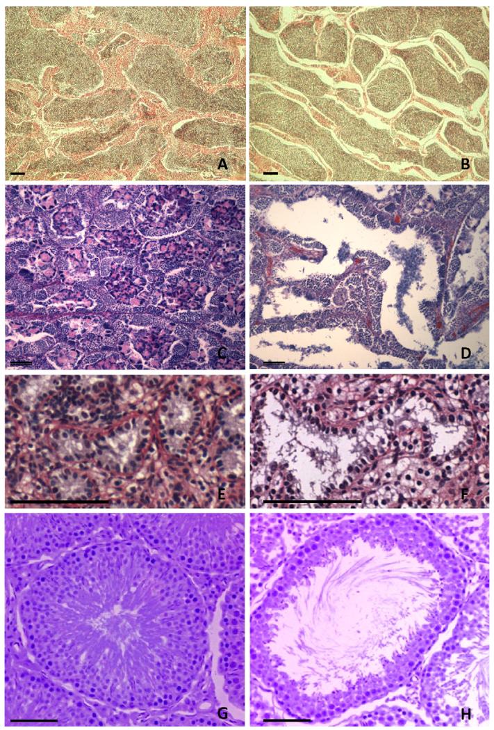
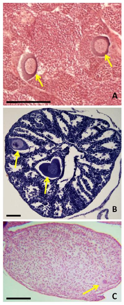
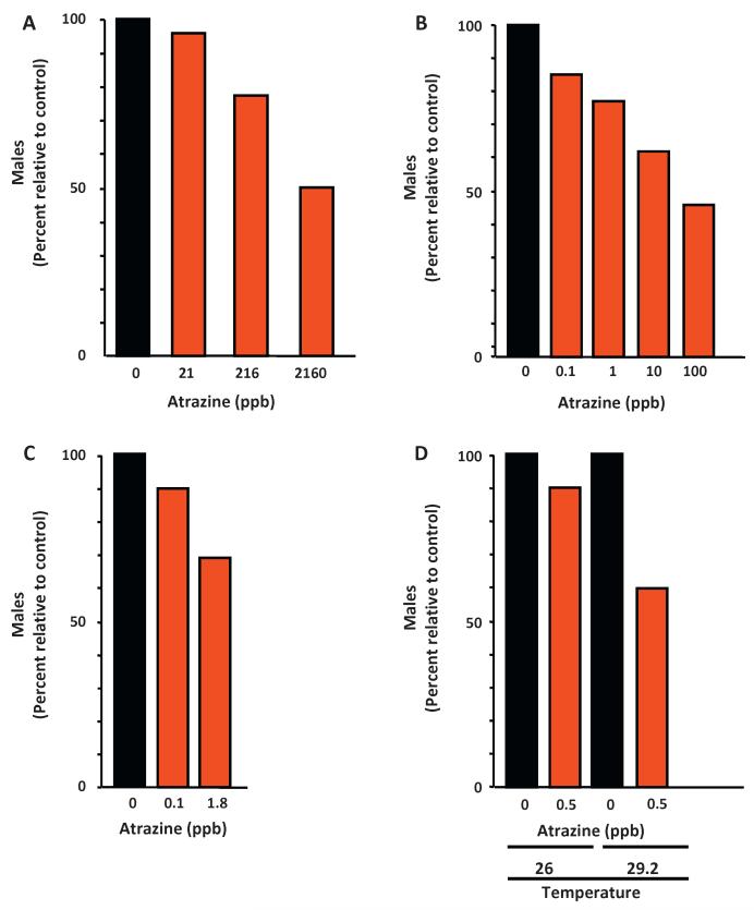
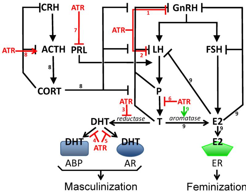
Atrazine is the most commonly detected pesticide contaminant of ground water, surface water, and precipitation. Atrazine is also an endocrine disruptor that, among other effects, alters male reproductive tissues when animals are exposed during development. Here, we apply the nine so-called "Hill criteria" (Strength, Consistency, Specificity, Temporality, Biological Gradient, Plausibility, Coherence, Experiment, and Analogy) for establishing cause-effect relationships to examine the evidence for atrazine as an endocrine disruptor that demasculinizes and feminizes the gonads of male vertebrates. We present experimental evidence that the effects of atrazine on male development are consistent across all vertebrate classes examined and we present a state of the art summary of the mechanisms by which atrazine acts as an endocrine disruptor to produce these effects. Atrazine demasculinizes male gonads producing testicular lesions associated with reduced germ cell numbers in teleost fish, amphibians, reptiles, and mammals, and induces partial and/or complete feminization in fish, amphibians, and reptiles. These effects are strong (statistically significant), consistent across vertebrate classes, and specific. Reductions in androgen levels and the induction of estrogen synthesis - demonstrated in fish, amphibians, reptiles, and mammals - represent plausible and coherent mechanisms that explain these effects. Biological gradients are observed in several of the cited studies, although threshold doses and patterns vary among species. Given that the effects on the male gonads described in all of these experimental studies occurred only after atrazine exposure, temporality is also met here. Thus the case for atrazine as an endocrine disruptor that demasculinizes and feminizes male vertebrates meets all nine of the "Hill criteria".


Figures

Fig. 1
Fig. 1
Atrazine-induced histologic lesions in testes of vertebrates. Histologic sections from a fish (A and B), an amphibian (C and D), a reptile (E and F) and a mammal (G and H) are shown. Histologic sections of testes of goldfish (Carassius auratus) controls (A) and after 21 days of exposure to water containing atrazine at 1,000 μg/L (B). Note the progressive increase in gaps in the interstitium between lobules. Sections were stained with Regaud’s hematoxylin, phloxine and light green. For details see Spano et al. [38]. Histological section of testes in African clawed frogs (Xenopus laevis) controls (C) and after exposure to atrazine at 2.5 μg/L throughout larval and postmetamorphic development (D). Sections were stained in Harris’ hematoxylin and eosin. For details see Hayes et al. [39]. Photomicrographs showing seminiferous tubules from a vehicle (E) and an atrazine-treated (F) caiman. Tissue sections were stained with Picrosirius solution and counterstained with Harris hematoxylin. For details, see Rey et al. [40]. Testicular tubules of control rats (G) and tubule of rats given atrazine at 200 mg/kg by gavage for 15 days (H). Atrazine-exposed rats were characterized by luminal dilation. Extended dosing up to 40 days resulted in testicular atrophy, which was mostly formed by Sertoli-only tubules (not shown). Sections were stained with hematoxylin and eosin. For details see Victor-Costa et al. [41]. Bar = 100 μ in all panels.
Fig. 2
Fig. 2
Partial feminization by atrazine in vertebrates. Testicular oocytes are induced by atrazine in fish (A), amphibians (B), and reptiles (C). Testes from an adult fathead minnow (Pimephales promelas) (A) exposed to water containing atrazine at 5 μg/L for 14 days presenting multiple testicular oocytes within the gametogenic and supportive cellular structures of the testes (Photo courtesy Diana Papoulias, U.S. Geological Survey, Columbia Environmental Research Center, Columbia, MO, USA). See Tillitt et al. [43] for details of experimental conditions. Testicular oocytes in the testes of a male Rana pipiens [B] exposed to atrazine at 0.1 μg/L. Section stained in Harris’ hematoxylin and eosin. For details see Hayes et al. [26]. Testicular oocyte (stained in Harris’ hematoxylin and eosin) in a snapping turtle (Chelydra serpentine) exposed to soil treated with atrazine at a typical application rate (3.1 L/ha). For details see de Solla et al. [47]. Bar = 100 μ in all panels.
Fig. 3
Fig. 3
Complete sex reversal by atrazine in vertebrates. Atrazine exposure causes a loss of males in exposed fish (A), amphibians (B and C), and reptiles (D). In fish, atrazine exposure produced a concentration-dependent decrease in the frequency of males [48]. (A) Similarly atrazine produced a concentration-dependent decrease in the frequency of males in African clawed frogs (Xenopus laevis) in the laboratory [49] (B) and in leopard frogs (Rana pipiens) exposed in semi-field conditions [50] (C). In red-eared sliders (Trachemys scripta elegans), atrazine seemed to reduce the number of males slightly at 26°, and a statistically significant reduction was evident at 29.2° [51] (D). In all cases, data are shown as percent males in experimental groups relative to controls. Figures A and B were adapted from Hayes et al. [39]. Original data for panel C from Langlois et al. [50] and original data were from panel D from Willingham et al. [51].
Fig. 4
Fig. 4
Multiple mechanisms of action have been identified for atrazine’s demasculinizing and feminizing effects on male gonads. Arrows indicate processes that are increased; bars indicate processes that are inhibited. Red lines indicate demasculinizing pathways that are directly affected by ATR and green lines indicate feminizing pathways that are directly affected by ATR. Numbers on pathways, refer to mechanisms listed in the text (see Section 1.5). ABP = androgen binding protein, ACTH = adrenocorticotropic hormone, AR = androgen receptor, ATR = atrazine, CORT = cortisol/corticosterone, CRH = corticotrophin-releasing hormone, DHT = dihydrotestosterone, E2 = 17β estradiol, ER = estrogen receptor, FSH = follicle stimulating hormone, GnRH = gonadotropin stimulating hormone, LH = luteinizing hormone, P = progesterone, PRL = prolactin., and T = testosterone (For interpretation of the references to colour in this figure legend, the reader is referred to the web version of the article.)

References

    1. Solomon K, et al. Ecological risk assessment of atrazine in North American surface waters. Environ. Toxicol. Chem. 1996;15:31–76. - PubMed
    1. Boyd R. Herbicides and herbicide degradates in shallow groundwater and the Cedar River near a municipal well field, Cedar Rapids, Iowa, Sci. Total Environ. 2000;248:241–253. - PubMed
    1. Capel P, Larson S. Effect of scale on the behavior of atrazine in surface waters. Environ. Sci. Technol. 2001;35(4):648–657. - PubMed
    1. Du Preez LH, et al. Seasonal exposures to triazine and other pesticides in surface waters in the western Highveld corn-production region in South Africa. Environ. Pollut. 2005;135(1):131–141. - PubMed
    1. Fenelon J, Moore R. Transport of agrichemicals to ground and surface waters in a small central Indiana watershed. J. Environ. Qual. 1998;27:884–894.

Publication types