. 2011 Apr;141(4):1020-8.
doi: 10.1016/j.jtcvs.2010.06.023.

The collateral network concept: a reassessment of the anatomy of spinal cord perfusion

Affiliations

The collateral network concept: a reassessment of the anatomy of spinal cord perfusion

Christian D Etz et al. J Thorac Cardiovasc Surg. 2011 Apr.

Abstract

Objective: Prevention of paraplegia after repair of thoracoabdominal aortic aneurysm requires understanding the anatomy and physiology of the spinal cord blood supply. Recent laboratory studies and clinical observations suggest that a robust collateral network must exist to explain preservation of spinal cord perfusion when segmental vessels are interrupted. An anatomic study was undertaken.

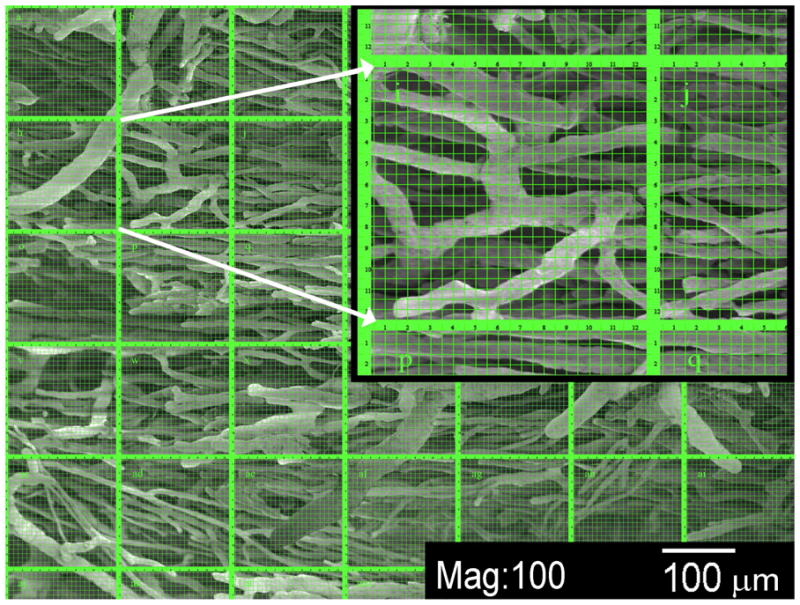
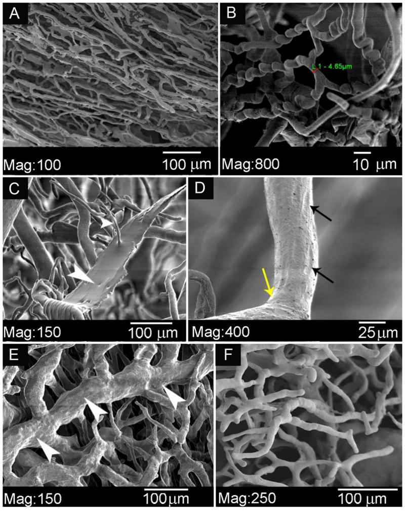
Methods: Twelve juvenile Yorkshire pigs underwent aortic cannulation and infusion of a low-viscosity acrylic resin at physiologic pressures. After curing of the resin and digestion of all organic tissue, the anatomy of the blood supply to the spinal cord was studied grossly and with light and electron microscopy.

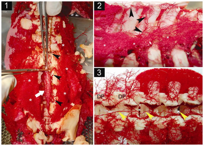
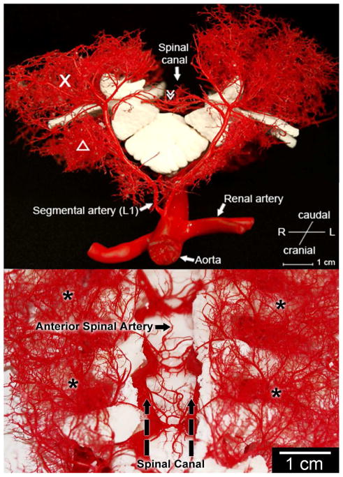
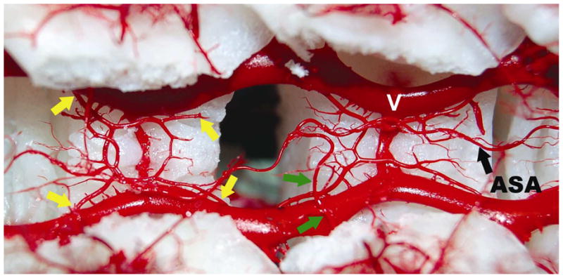
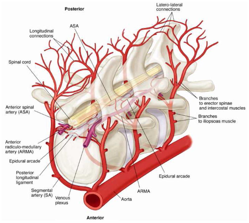
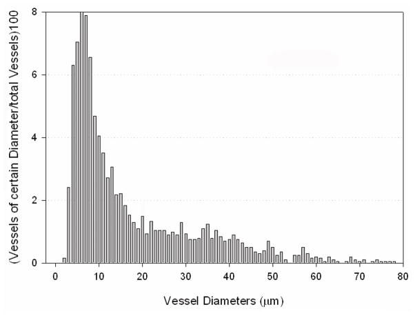
Results: All vascular structures at least 8 μm in diameter were preserved. Thoracic and lumbar segmental arteries give rise not only to the anterior spinal artery but to an extensive paraspinous network feeding the erector spinae, iliopsoas, and associated muscles. The anterior spinal artery, mean diameter 134 ± 20 μm, is connected at multiple points to repetitive circular epidural arteries with mean diameters of 150 ± 26 μm. The capacity of the paraspinous muscular network is 25-fold the capacity of the circular epidural arterial network and anterior spinal artery combined. Extensive arterial collateralization is apparent between the intraspinal and paraspinous networks, and within each network. Only 75% of all segmental arteries provide direct anterior spinal artery-supplying branches.

Conclusions: The anterior spinal artery is only one component of an extensive paraspinous and intraspinal collateral vascular network. This network provides an anatomic explanation of the physiological resiliency of spinal cord perfusion when segmental arteries are sacrificed during thoracoabdominal aortic aneurysm repair.


Figures

Figure 1
Figure 1. Processing of the acrylic cast
1.1 After soft tissue maceration and multiple cleaning steps using distilled water, the spinal canal (black arrows) is opened dorsally by cutting the pedicles of the vertebrae. The casts of spinal cord arteries (white arrow: lower lumber cord) are dissected and freed from neural and glial tissues. Asterisks: lower paraspinous musculature. 1.2: Lateral view of casts of vessels along dorsal processes show paravertebral extramuscular arcades consisting of arterioles that interconnect segmental levels longitudinally. *extensive vasculature of paraspinous muscles. 1.3: Dorsal view of the opened spinal canal onto the lower thoracic (left) and upper lumbar (right) segments. Yellow arrows show the anterior spinal artery. DP: Distribution points of single dorsal segmental arteries, where the dorsal mainstem divides into an extensive muscular paraspinous vascular tree, giving rise to the different intraspinal branches.
Figure 2
Figure 2. Anatomy of the collateral network : sagittal (A) and dorsal (B) view
The macroscopic appearance of casts of a pair of dorsal segmental vessels at L1. The dorsal process is removed. X designates the paraspinous muscular vasculature providing extensive longitudinal arterio-arteriolar connections. Δ iliopsoas muscle. ≫ Anterior spinal artery.
Figure 3
Figure 3. Relationship of anterior spinal artery (ASA) and repetitive epidural arcades
Dorsal view into the opened spinal canal showing the dorsal surface of two vertebral bodies. The spinal cord is removed to clarify the anatomic location of the epidural circular arcades and anterior spinal artery (ASA). V: epidural venous plexus. Anterior to the extensive venous plexus, four arteriolar branches (yellow arrows) contribute to one circular epidural arcade. This pattern is repeated at the level of each vertebral segment. These vascular structures connect the segments side-to-side as well as longitudinally. They connect with the main stems of the segmental arteries, and can therefore be considered to contribute indirectly to the ASA. Green arrows designate the anterior radiculo-medullary artery, which connects directly with the anterior spinal artery.
Figure 4
Figure 4. Schematic Diagram of the Blood Supply to the Spinal Cord
Schematic diagram demonstrating the relationships, relative sizes and interconnections among the segmental arteries (SA), the anterior radiculomedullary arteries (ARMA) the epidural arcades, and the anterior spinal artery (ASA). Longitudinal anastomoses along the dorsal processes of the spine as well as dorsal communications (interstitial connections) between right and left branches of the SA are also shown.
Figure 5
Figure 5. Cast analysis using scanning electron microscopy
SEM of paraspinous vascular cast specimens after soft tissue maceration and multiple cleaning steps. The image shows an arteriolar intramuscular network, and the counting grid used for vessel diameter distribution analysis within the collateral network is superimposed upon it. The inset shows detail of a single counting grid, one of a total of 35 per image. The vessels shown have diameters of small precapillary arterioles down to 15 – 20 μm.
Figure 6
Figure 6. Microanatomy of the collateral network
An overview of casts of paraspinous vascular structures. A : A capillary micromesh. B : Detail of typical «corkscrew- shaped» capillaries. C: Artifact of acrylic resin which has leaked through a damaged vessel wall. D : Endothelial nuclear imprints on a precapillary arteriole. E : A network of venules, in comparison to F, an arteriolar network.
Figure 7
Figure 7. Size distribution of capillary and arterioles within the paraspinous collateral network
The graph shows the distribution of vessels of different diameters (up to 80 μm) within the paraspinous vascular network. The number of times a vessel of a certain diameter was measured is shown as a percentage of the number of all analyzed vessels (n = 2030 total).

Similar articles

Cited by

References

    1. Heinemann MK, Brassel F, Herzog T, Dresler C, Becker H, Borst HG. The role of spinal angiography in operations on the thoracic aorta: myth or reality? Ann Thorac Surg. 1998;65(2):346–51. - PubMed
    1. Kieffer E, Richard T, Chiras J, Godet G, Cormier E. Preoperative spinal cord arteriography in aneurysmal disease of the descending thoracic and thoracoabdominal aorta: preliminary results in 45 patients. Ann Vasc Surg. 1989;3(1):34–46. - PubMed
    1. Williams GM, Perler BA, Burdick JF, Osterman FA, Jr, Mitchell S, Merine D, et al. Angiographic localization of spinal cord blood supply and its relationship to postoperative paraplegia. J Vasc Surg. 1991;13(1):23–33. discussion -5. - PubMed
    1. Williams GM, Roseborough GS, Webb TH, Perler BA, Krosnick T. Preoperative selective intercostal angiography in patients undergoing thoracoabdominal aneurysm repair. J Vasc Surg. 2004;39(2):314–21. - PubMed
    1. Kawaharada N, Morishita K, Fukada J, Yamada A, Muraki S, Hyodoh H, et al. Thoracoabdominal or descending aortic aneurysm repair after preoperative demonstration of the Adamkiewicz artery by magnetic resonance angiography. Eur J Cardiothorac Surg. 2002;21(6):970–4. - PubMed

Publication types