. 2011 Apr;141(4):1029-36.
doi: 10.1016/j.jtcvs.2010.06.017.

The collateral network concept: remodeling of the arterial collateral network after experimental segmental artery sacrifice

Affiliations

The collateral network concept: remodeling of the arterial collateral network after experimental segmental artery sacrifice

Christian D Etz et al. J Thorac Cardiovasc Surg. 2011 Apr.

Abstract

Objective: A comprehensive strategy to prevent paraplegia after open surgical or endovascular repair of thoracoabdominal aortic aneurysms requires a thorough understanding of the response of the collateral network to extensive segmental artery sacrifice.

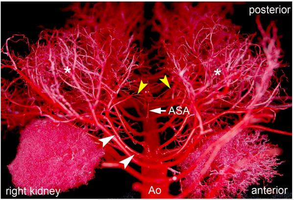
Methods: Ten Yorkshire pigs underwent perfusion with a low-viscosity acrylic resin. With the use of cardiopulmonary bypass, 2 animals each were perfused in the native state and immediately, 6 hours, 24 hours, and 5 days after sacrifice of all segmental arteries (T4-L5). After digestion of surrounding tissue, the vascular cast of the collateral network underwent analysis of arterial and arteriolar diameters and the density and spatial orientation of the vasculature using light and scanning electron microscopy.

Results: Within 24 hours, the diameter of the anterior spinal artery had increased significantly, and within 5 days the anterior spinal artery and the epidural arterial network had enlarged in diameter by 80% to 100% (P < .0001). By 5 days, the density of the intramuscular paraspinous vessels had increased (P < .0001), a shift of size distribution from small to larger arterioles was seen (P = .0002), and a significant realignment of arterioles parallel to the spinal cord had occurred (P = .0005).

Conclusions: Within 5 days after segmental artery occlusion, profound anatomic alterations in the intraspinal and paraspinous arteries and arterioles occurred, providing the anatomic substrate for preservation of spinal cord blood flow via collateral pathways.


Figures

Figure 1
Figure 1
Vascular cast of the abdominal aorta (Ao), renal arteries, kidneys and dorsal lumbar segmental arteries in a Yorkshire pig (cranio-caudal view, segment L1 in foreground). Soft tissue and some bony structures have been completely removed in order to demonstrate the distribution and branching of dorsal segmental arteries. White arrowheads show the main trunks of the segmental arteries following the contour of the removed vertebral bodies. Note the extensive contribution of the segmental arteries to the paraspinous musculature (asterisks). Two to three branches of each segmental artery trunk reach the intraspinal collateral network (yellow arrowheads) as well as the anterior spinal artery (ASA).
Figure 2
Figure 2
Increase in the diameter of the anterior spinal artery diameter 24 hours and 5 days after occlusion of all segmental arteries in the pig model. ASA: Anterior spinal artery. SA: Segmental artery. Native: patent segmental inflow into collateral networks. Separate measurements of the upper thoracic, lower thoracic and lumbar segments have been merged.
Figure 3
Figure 3
Epidural arcade diameter changes at 24 and 120 hours after complete segmental artery ligation in the pig model.
Figure 4
Figure 4
Increase in diameters of epidural arcades (left, black arrowheads) and anterior spinal artery (right, black arrowheads) in the native pig (Nat) as compared to a pig perfused five days after extensive segmental artery occlusion (120 h). Soft tissue was removed completely from the samples, but the bony vertebral column was preserved. Note the specific enlargement of the longitudinally oriented vessels of the epidural arcade on the left.
Figure 5
Figure 5
Paraspinous vessel density change 120 hours after complete segmental artery ligation in the pig. Bars represent the percentage of randomly determined coordinates which included a blood vessel.
Figure 6
Figure 6
Shift in vessel diameter distribution in the paraspinous network 120 hours after complete segmental artery ligation in the pig. In the lower thoracic and lumbar regions, a clear shift from 1-10μm vessels (capillaries) to 10-30μm vessels (small arterioles) is apparent.
Figure 7
Figure 7
Progressive parallelization of small vessels in the paraspinal network 24 and 120 hours after segmental artery ligation in the pig. Representative details from scanning electron microscopy images of the paraspinous vascular system show the change in vessel orientation after occlusion of segmental inflow into the spinal collateral networks. Insets show the calculated deviation of measured vessel angles for the images. Top: native animal with patent segmental inflow. Mid: animal 24 hours after segmental artery occlusion. Bottom: animal five days after segmental artery occlusion. All images were taken from low thoracic and high lumbar segments.
Figure 8
Figure 8
Extent of change in spatial orientation of paraspinous microvessels (arterioles up to 40 micrometers) 5 days following complete segmental artery occlusion (T4-L5) as compared to native pigs with patent segmental inflow into the collateral networks. For every segmental level, the mean orientation of all analyzed vessels was calculated. The graph shows the mean values of deviation from this calculated orientation: a random orientation of analyzed vessels results in higher values (45 degrees). Values closer to zero represent a vascular network with a unidirectional pattern. The images on the left show examples of both extremes: (lower image: native vascular network; upper image: vascular network five days after segmental artery occlusion). Note that the biggest difference in vascular orientation was found in the lower thoracic segments.

References

    1. Maniar HS, Sundt TM, 3rd, Prasad SM, Chu CM, Camillo CJ, Moon MR, et al. Delayed paraplegia after thoracic and thoracoabdominal aneurysm repair: a continuing risk. Ann Thorac Surg. 2003;75(1):113–9. discussions 19-20. - PubMed
    1. Etz CD, Luehr M, Kari FA, Bodian CA, Smego D, Plestis KA, et al. Paraplegia after extensive thoracic and thoracoabdominal aortic aneurysm repair: does critical spinal cord ischemia occur postoperatively? J Thorac Cardiovasc Surg. 2008;135(2):324–30. - PubMed
    1. Etz CD, Halstead JC, Spielvogel D, Shahani R, Lazala R, Homann TM, et al. Thoracic and thoracoabdominal aneurysm repair: is reimplantation of spinal cord arteries a waste of time? Ann Thorac Surg. 2006;82(5):1670–7. - PubMed
    1. Etz CD, Di Luozzo G, Zoli S, Lazala R, Plestis KA, Bodian CA, et al. Direct spinal cord perfusion pressure monitoring in extensive distal aortic aneurysm repair. Ann Thorac Surg. 2009;87(6):1764–73. discussion 73-4. - PubMed
    1. Etz CD, Kari FA, Mueller CS, Silovitz D, Brenner R, Lin HM, Griepp RB. The collateral network concept: a reassessment of the anatomy of spinal cord perfusion. J Thorac Cardiovasc Surg. 2010 In preparation. - PMC - PubMed

Publication types