Review
. 2011 Apr;157(4):200-8.
doi: 10.1016/j.trsl.2011.01.001. Epub 2011 Feb 4.

Involvement of microRNAs in lung cancer biology and therapy

Affiliations
Review

Involvement of microRNAs in lung cancer biology and therapy

Xi Liu et al. Transl Res. 2011 Apr.

Abstract

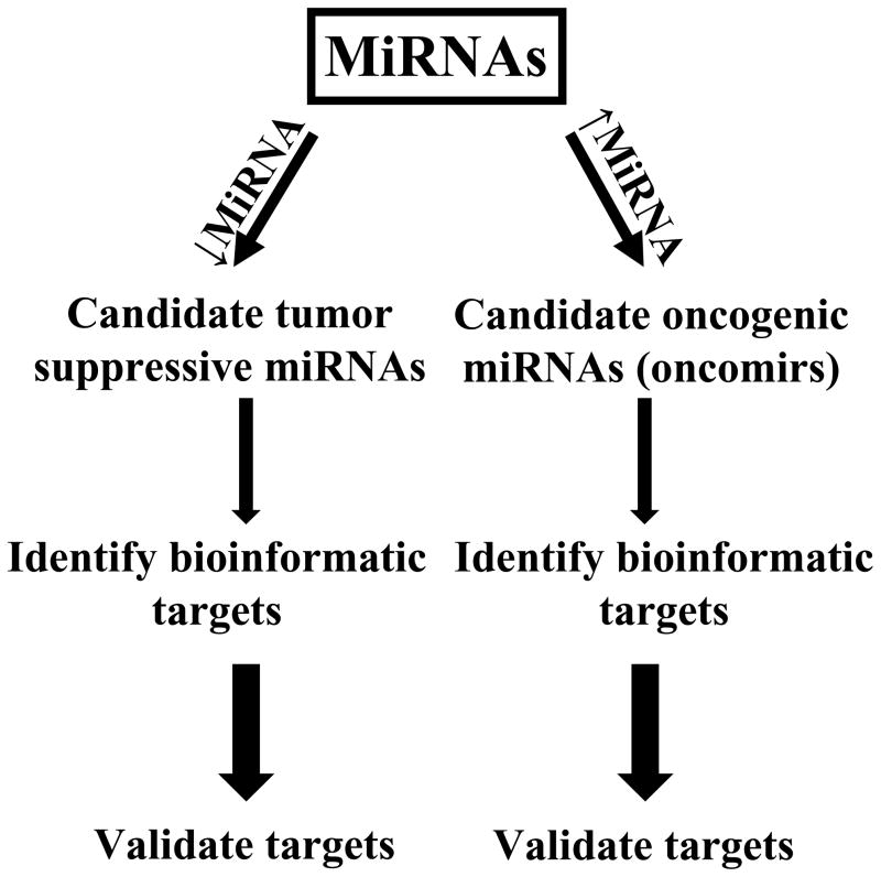
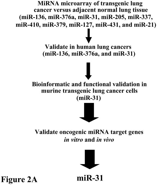
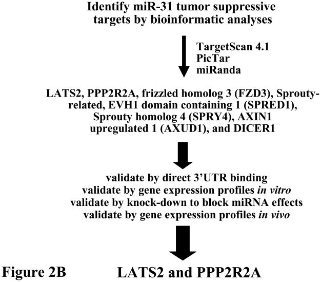
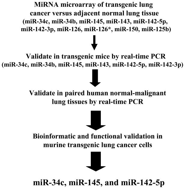
MicroRNAs (miRNAs) are a class of small RNAs that regulate gene expression. Expression profiles of specific miRNAs have improved cancer diagnosis and classification as well as provided prognostic information in many human cancers, including lung cancer. Tumor-suppressive and oncogenic miRNAs were uncovered in lung carcinogenesis. The biological functions of these miRNAs in lung cancer were validated recently in well-characterized cellular, murine transgenic as well as transplantable lung cancer models, and in human paired normal-malignant lung tissue banks and tissue arrays. Tumor-suppressive and oncogenic miRNAs that were identified in lung cancer will be reviewed here. Emphasis is placed on highlighting those functionally validated miRNAs that are not only biomarkers of lung carcinogenesis but also candidate pharmacologic targets. How these miRNA findings advance an understanding of lung cancer biology and how they could improve lung cancer therapy are discussed in this article.


Figures

Figure 1
Figure 1
Identification and functional characterization of oncogenic and tumor suppressive miRNAs in lung cancer. Candidate tumor suppressive miRNAs are repressed (↓miRNA) while candidate oncogenic (oncomir) miRNAs are increased (↑miRNA) in their expression within lung cancers (relative to adjacent normal lung tissues). Bioinformatic analysis is used to prioritize potential target genes before functional validation, as described in the text. The increased arrow sizes are meant to convey an increased stringency of selection for the highlighted miRNAs.
Figure 2
Figure 2
Comprehensive analysis of known miRNAs was performed to uncover those that were increased in lung cancers relative to adjacent normal lung. (A) Initial analysis was performed in transgenic cyclin E-driven mouse models that recapitulated frequent features of human lung cancer such as presence of chromosome instability, hedgehog pathway activation, single or multiple pre-malignant and malignant (adenocarcinoma) lung lesions and even the onset of metastases. Results were confirmed and extended for a subset of highlighted miRNAs (see figure) within a panel of paired normal-malignant human lung tissues. Gain and loss of function studies were performed in murine and human lung cancer cells to explore the anti-neoplastic effects of each highlighted miRNA. This led to finding miR-31 as a key oncomir in the lung. This conclusion was made based on engineered loss of miR-31 that caused a significant reduction in lung cancer formation after transplantation of these cells into syngeneic mice, as discussed in the text. (B) Bioinformatic analysis was performed using the indicated programs shown in this panel. This uncovered the tumor suppressors (LATS2 and PPP2R2A) as regulators of the oncogenic effects of miR-31. The increase in arrow size conveys the increased stringency of selection for the highlighted miRNAs.
Figure 2
Figure 2
Comprehensive analysis of known miRNAs was performed to uncover those that were increased in lung cancers relative to adjacent normal lung. (A) Initial analysis was performed in transgenic cyclin E-driven mouse models that recapitulated frequent features of human lung cancer such as presence of chromosome instability, hedgehog pathway activation, single or multiple pre-malignant and malignant (adenocarcinoma) lung lesions and even the onset of metastases. Results were confirmed and extended for a subset of highlighted miRNAs (see figure) within a panel of paired normal-malignant human lung tissues. Gain and loss of function studies were performed in murine and human lung cancer cells to explore the anti-neoplastic effects of each highlighted miRNA. This led to finding miR-31 as a key oncomir in the lung. This conclusion was made based on engineered loss of miR-31 that caused a significant reduction in lung cancer formation after transplantation of these cells into syngeneic mice, as discussed in the text. (B) Bioinformatic analysis was performed using the indicated programs shown in this panel. This uncovered the tumor suppressors (LATS2 and PPP2R2A) as regulators of the oncogenic effects of miR-31. The increase in arrow size conveys the increased stringency of selection for the highlighted miRNAs.
Figure 3
Figure 3
Comprehensive analysis of known miRNAs was performed to uncover those that were repressed in murine lung cancers relative to adjacent normal lung tissues. Results were confirmed and extended for a subset of highlighted miRNAs in a panel of paired normal-malignant human lung tissues. Gain and loss of function studies were independently performed in murine and human lung cancer cells to explore the anti-neoplastic effects of each highlighted miRNA. Of these species, three prominent growth suppressive miRNAs were found and were each functionally validated. These were: miR-34c, miR-145, and miR-142-5p. The increase in arrow size conveys the increased stringency of selection for the highlighted miRNAs.
Figure 4
Figure 4
Spatial characterization of miRNA expression profiles in murine and human lung tissues. Representative in situ hybridization (ISH) assay results are displayed using locked nucleic acid (LNA)-modified probes against candidate tumor suppressive or oncogenic miRNAs. These assays were performed using formalin-fixed paraffin-embedded normal or malignant lung tissues, as previously described., The upper panel shows the miRNA expression patterns of the indicated miRNAs in age-matched non-transgenic sibling controls (normal lung tissue) and in a cyclin E-driven mouse model of lung cancer, as previously described. ISH analyses indicated the predominant bronchial epithelial cell expression pattern for miR-34c (arrows) as well as for expression of miR-145 in epithelial cells (small arrows), though miR-145 was also expressed in the smooth muscle cells of the lamina muscularis of the mucosa and vasculature (arrows). In contrast, miR-126 expression was expressed in endothelial cells (arrows) and by this profile was viewed as having a lower priority for miRNA functional assessment than either miR-34c or miR-145. The lower panel displays the ISH expression patterns for miR-31 and for the 18S ribosomal RNA (as control for RNA integrity) in paired human normal lung tissue versus adjacent lung cancer (adenocarcinoma). This ISH analysis confirmed augmented miR-31 expression (relative to adjacent normal lung) within both murine and human lung cancers. Representative hematoxylin and eosin (H & E) stained lung tissues are displayed.

References

    1. Lagos-Quintana M, Rauhut R, Lendeckel W, Tuschl T. Identification of novel genes coding for small expressed RNAs. Science. 2001;294:853–8. - PubMed
    1. Lau NC, Lim LP, Weinstein EG, Bartel DP. An abundant class of tiny RNAs with probable regulatory roles in Caenorhabditis elegans. Science. 2001;294:858–62. - PubMed
    1. Lee RC, Ambros V. An extensive class of small RNAs in Caenorhabditis elegans. Science. 2001;294:862–4. - PubMed
    1. Kim VN. MicroRNA biogenesis: coordinated cropping and dicing. Nat Rev Mol Cell Biol. 2005;6:376–85. - PubMed
    1. Yi R, Qin Y, Macara IG, Cullen BR. Exportin-5 mediates the nuclear export of pre-microRNAs and short hairpin RNAs. Genes Dev. 2003;17:3011–6. - PMC - PubMed

Publication types