. 2011 Jun 3;409(2):159-76.
doi: 10.1016/j.jmb.2011.03.026. Epub 2011 Mar 21.

The double-length tyrosyl-tRNA synthetase from the eukaryote Leishmania major forms an intrinsically asymmetric pseudo-dimer

Affiliations

The double-length tyrosyl-tRNA synthetase from the eukaryote Leishmania major forms an intrinsically asymmetric pseudo-dimer

Eric T Larson et al. J Mol Biol. .

Abstract

The single tyrosyl-tRNA synthetase (TyrRS) gene in trypanosomatid genomes codes for a protein that is twice the length of TyrRS from virtually all other organisms. Each half of the double-length TyrRS contains a catalytic domain and an anticodon-binding domain; however, the two halves retain only 17% sequence identity to each other. The structural and functional consequences of this duplication and divergence are unclear. TyrRS normally forms a homodimer in which the active site of one monomer pairs with the anticodon-binding domain from the other. However, crystal structures of Leishmania major TyrRS show that, instead, the two halves of a single molecule form a pseudo-dimer resembling the canonical TyrRS dimer. Curiously, the C-terminal copy of the catalytic domain has lost the catalytically important HIGH and KMSKS motifs characteristic of class I aminoacyl-tRNA synthetases. Thus, the pseudo-dimer contains only one functional active site (contributed by the N-terminal half) and only one functional anticodon recognition site (contributed by the C-terminal half). Despite biochemical evidence for negative cooperativity between the two active sites of the usual TyrRS homodimer, previous structures have captured a crystallographically-imposed symmetric state. As the L. major TyrRS pseudo-dimer is inherently asymmetric, conformational variations observed near the active site may be relevant to understanding how the state of a single active site is communicated across the dimer interface. Furthermore, substantial differences between trypanosomal TyrRS and human homologs are promising for the design of inhibitors that selectively target the parasite enzyme.


Figures

Figure 1
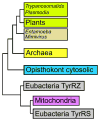
Figure 1. Phylogenetic relationship of TyrRS sequences
Cartoon representation of the phylogenetic relationship of TyrRS sequences from trypanosomatids, Mimivirus, plants, opisthokont cytosolic homologs, mitochondrial homologs, archaea, and bacteria. The cartoon combines information from previously published work,,.
Figure 2
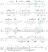
Figure 2. Gene structure and motifs in double-length TyrRS homologs, other eukaryotes and archaea
The coloring of the catalytic (brown) and anticodon-binding (blue) domains corresponds to that of the L. major TyrRS structure shown in Figure 3. The HIGH and KMSKS active site motifs are common to the Class I catalytic domain. The AIDQ motif is characteristic of the ATP-binding site in TyrRS and TrpRS. The AC1 motif (KIKKAYS in L. major TyrRS) corresponds to the residues in helix α11 of the eukary-otic/archaeal TyrRS anticodon-binding domain that interact with the anticodon stem of tRNATyr. The AC2 motif (LHPADLK in L. major TyrRS) contains the residues in helix α14 that specifically recognize the anticodon bases G34 and U/ψ35.
Figure 3
Figure 3. Pseudo-dimeric L. major TyrRS and true dimeric human TyrRS
The inherently asymmetric L. major pseudo-dimer shown is that of the tyrosinol:TyrRS complex (PDBID 3p0i). The human dimer is perfectly symmetric as seen in the crystal structure of the two core TyrRS domains in complex with tyrosinol (PDBID 1q11). The catalytic domain is colored brown in both structures, dark brown for the N-terminal (functional) copy in L. major TyrRS, light brown for the C-terminal (presumed nonfunctional) copy in L. major TyrRS, intermediate brown in both monomers of the human TyrRS. The anticodon-binding domain is colored blue in the L. major TyrRS, dark blue for the N-terminal copy and light blue for the C-terminal (functional) copy. The anticodon-binding domain is colored yellow in both monomers of human TyrRS. The linker residues connecting the N- and C-terminal halves of the L. major TyrRS are shown in magenta; the analogous residues residues in human TyrRS are also shown in magenta in the second monomer. Crystallographically observed bound tyrosinol molecules in both structures are shown in green. Figure generated using PyMOL.
Figure 4
Figure 4. Sequence alignment and secondary structure of L. major and human cytosolic TyrRS
Structure-based alignment of the N- and C-terminal halves of the L. major TyrRS sequence together with the sequence of the core domains of human cytosolic TyrRS. Secondary structure elements for the N-terminal half of L. major TyrRS from the cubic form of the tyrosinol complex are shown above the sequences, except that the extended part of the longer α5 helix seen in the fisetin complex is shown in green (residues 84–88). Secondary structure elements for the C-terminal half of L. major TyrRS are shown underneath the sequences. Blue boxes indicate the canonical Class Ic catalytic domain motifs. Red boxes labeled AC1 and AC2 indicate the regions which are observed to interact with the tRNA anticodon stem in the yeast TyrRS:tyrosyladenylate:tRNATyr complex (PDBID 2dlc). Residues of the human sequence that are within 4 Å of the superimposed tyrosyladenylate molecule from the yeast complex are highlighted in cyan. Residues coordinating a K+ ion in the human structure are highlighted in magenta. Figure generated using TEXshade.
Figure 5
Figure 5. Two conformational states of the active site are observed in L. major TyrRS structures
The closed state of active site in the N-terminal half of L. major TyrRS as seen in the tyrosinol complex is shown in green. This state is characterized by a well-ordered 148–149 loop and a ‘curled’ conformation of the α4–α5 loop. The open state of active site as seen in the fisetin complex is shown in yellow. In this state, the 148–149 loop is disordered and the α4–α5 loop is present in an ‘extended’ conformation that is in contact with helix α7′ across the “pseudo-dimer” interface (shown in cyan). This is analogous to the active site of one monomer from a canonical TyrRS dimer establishing contact with the second monomer across the dimer interface.
Figure 6
Figure 6. Active site of L. major TyrRS; with electron density for ligands, and highlighting substantial sequence divergence from human TyrRSs
(a) Active site of L. major TyrRS with bound tyrosinol. Electron density is contoured at 4σ in an (mFobsFcalc) difference map with tyrosinol omitted from Fcalc. (b) Active site of L. major TyrRS with bound fisetin. Electron density is contoured at 4σ in an (mFobs – Fcalc) difference map with fisetin omitted from Fcalc. (c) Superposition of L. major TyrRS complexes with tyrosinol (yellow; PDBID 3p0h), fisetin (cyan; PDBID 3p0i), and yeast TyrRS with tyrosyladenylate (green; PDBID 2dlc). (d) Stereo view of the same superimposed ligands from panel c in the L. major TyrRS active site pocket, shown as a transparent surface so the rather buried ligands are not mostly concealed. Residues within 8 Å of the superimposed tyrosyladeny-late are colored to highlight the substantial differences between the L. major enzyme and the two human homologs. Identical residues are light green, those that differ from the mitochondrial enzyme are yellow, those that differ from the cytoplasmic enzyme are red, and those that differ from both human enzymes are orange. The view into the pocket is approximately 50° counterclockwise around the Y-axis with respect to the orientation in Figure 3.
Figure 7
Figure 7. Comparison of anticodon recognition regions of the N- and C-terminal halves of L. major TyrRS and yeast TyrRS
The N-terminal (dark blue) and C-terminal (light blue) copies of the L. major anticodon-binding domain are shown superimposed onto the yeast TyrRS:tRNATyr complex (PDBID 2dlc) (yellow). The three anticodon bases are labeled. The C-terminal region is more structurally similar that of the yeast enzyme than either are to that of the N-terminal region.
Figure 8
Figure 8. Thermal denaturation curves
(a) Thermal denaturation curves for L. major TyrRS alone and in the presence of tyrosine, ATP, tyrosine and ATP both, or fisetin. (b) Thermal denaturation curves for T. cruzi TyrRS alone and in the presence of tyrosinol, ATP, or tyrosinol and ATP both.
Figure 8
Figure 8. Thermal denaturation curves
(a) Thermal denaturation curves for L. major TyrRS alone and in the presence of tyrosine, ATP, tyrosine and ATP both, or fisetin. (b) Thermal denaturation curves for T. cruzi TyrRS alone and in the presence of tyrosinol, ATP, or tyrosinol and ATP both.

Similar articles

Cited by

References

    1. Eriani G, Delarue M, Poch O, Gangloff J, Moras D. Partition of tRNA synthetases into two classes based on mutually exclusive sets of sequence motifs. Nature. 1990;347:203–6. - PubMed
    1. Cusack S, Berthet-Colominas C, Hartlein M, Nassar N, Leberman R. A second class of synthetase structure revealed by X-ray analysis of Escherichia coli seryl-tRNA synthetase at 2.5 Å. Nature. 1990;347:249–255. - PubMed
    1. Wolf YI, Aravind L, Grishin NV, Koonin EV. Evolution of aminoacyl-tRNA synthetases–analysis of unique domain architectures and phylogenetic trees reveals a complex history of horizontal gene transfer events. Genome Res. 1999;9:689–710. - PubMed
    1. Brindefalk B, Viklund J, Larsson D, Thollesson M, Andersson SGE. Origin and evolution of the mitochondrial aminoacyl-tRNA synthetases. Mol Biol Evol. 2007;24:743–56. - PubMed
    1. Brown J, Robb F, Weiss R, Doolittle W. Evidence for the early divergence of tryptophanyl- and tyrosyl-tRNA synthetases. J Molec Evol. 1997;45:9–16. - PubMed