Review
. 2011 Jun;7(6):2355-73.
doi: 10.1016/j.actbio.2011.03.016. Epub 2011 Mar 21.

Bioactive glass in tissue engineering

Affiliations
Review

Bioactive glass in tissue engineering

Mohamed N Rahaman et al. Acta Biomater. 2011 Jun.

Abstract

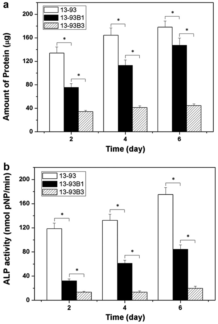
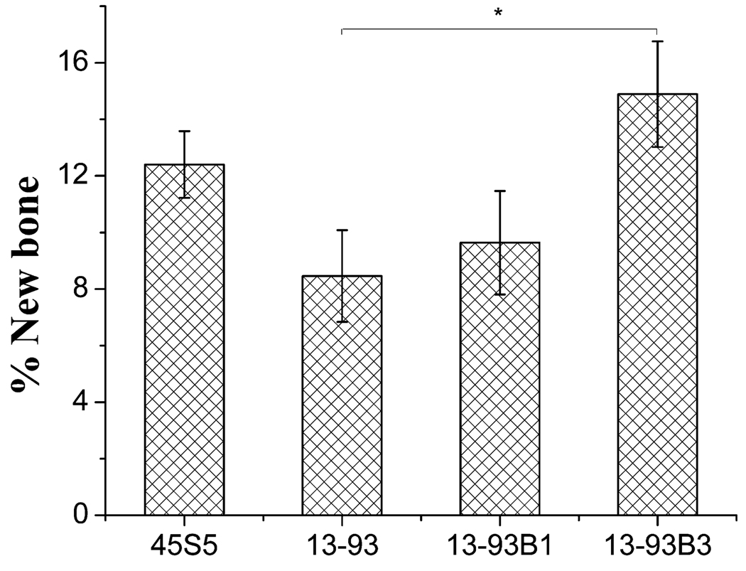
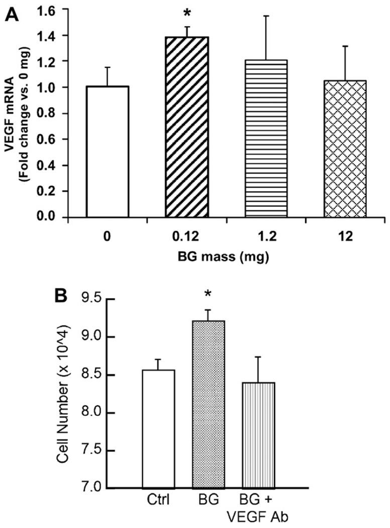
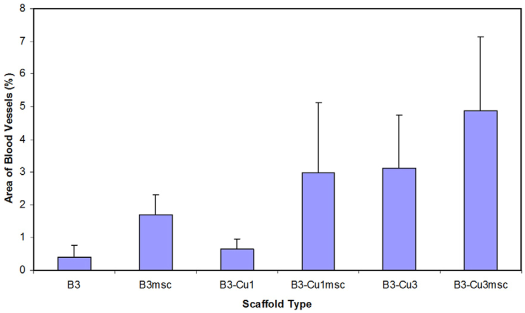
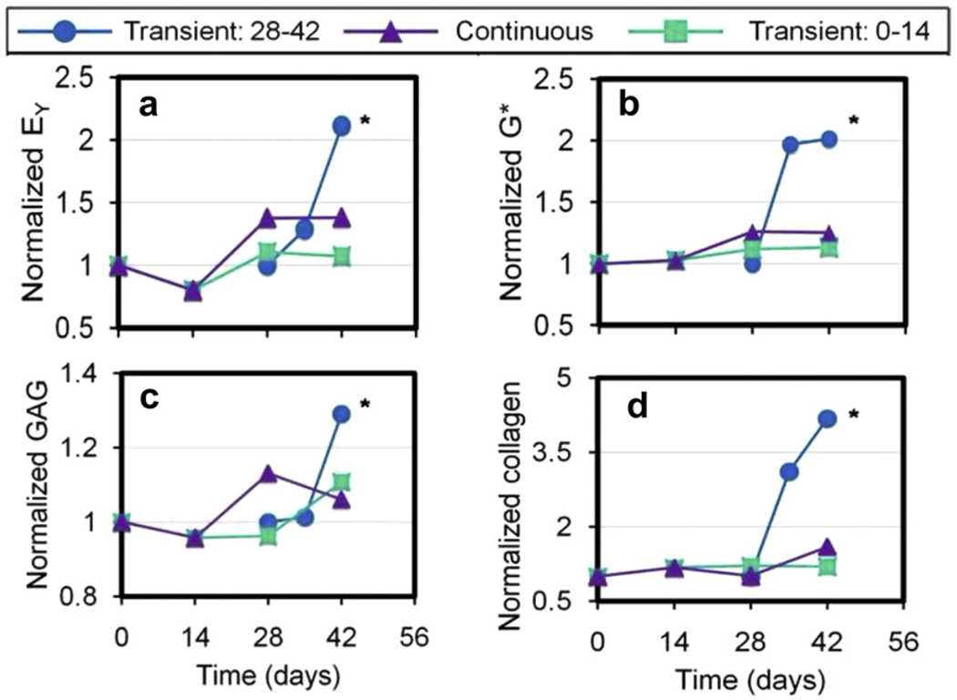
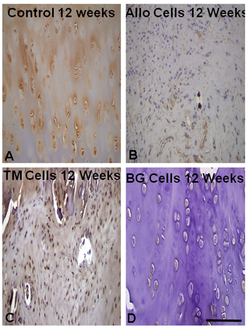
This review focuses on recent advances in the development and use of bioactive glass for tissue engineering applications. Despite its inherent brittleness, bioactive glass has several appealing characteristics as a scaffold material for bone tissue engineering. New bioactive glasses based on borate and borosilicate compositions have shown the ability to enhance new bone formation when compared to silicate bioactive glass. Borate-based bioactive glasses also have controllable degradation rates, so the degradation of the bioactive glass implant can be more closely matched to the rate of new bone formation. Bioactive glasses can be doped with trace quantities of elements such as Cu, Zn and Sr, which are known to be beneficial for healthy bone growth. In addition to the new bioactive glasses, recent advances in biomaterials processing have resulted in the creation of scaffold architectures with a range of mechanical properties suitable for the substitution of loaded as well as non-loaded bone. While bioactive glass has been extensively investigated for bone repair, there has been relatively little research on the application of bioactive glass to the repair of soft tissues. However, recent work has shown the ability of bioactive glass to promote angiogenesis, which is critical to numerous applications in tissue regeneration, such as neovascularization for bone regeneration and the healing of soft tissue wounds. Bioactive glass has also been shown to enhance neocartilage formation during in vitro culture of chondrocyte-seeded hydrogels, and to serve as a subchondral substrate for tissue-engineered osteochondral constructs. Methods used to manipulate the structure and performance of bioactive glass in these tissue engineering applications are analyzed.


Figures

Fig. 1
Fig. 1
Number of papers published per year in the field of “bioactive glass” (compiled from a literature search in Web of Science carried out in December 2010).
Fig. 2
Fig. 2
Microstructures of bioactive glass scaffolds created by a variety of processing methods: (a) thermal bonding (sintering) of particles (microspheres); (b) thermal bonding of short fibers; (c) “trabecular” microstructure prepared by a polymer foam replication technique; (d) oriented microstructure prepared by unidirectional freezing of suspensions (plane perpendicular to the orientation direction); (e) X-ray microCT image of the oriented scaffold shown in (d); (f) grid-like microstructure prepared by robocasting. Glass composition: (a) 16CaO−21Li2O−63B2O3; (b–e) 13–93; (f) 6P53B.
Fig. 3
Fig. 3
SEM images of nanofibrous bioactive glass: (a) borosilicate 13–93B1 glass with diameters in the range 150–450 nm prepared by electrospinning of a precursor solution (courtesy of C. Gao; Shinshu University, Japan); (b) bioactive glass with diameters in the range 100–800 nm, prepared from a melt-derived glass (courtesy of Mo-Sci Corp., Rolla, MO, USA).
Fig. 4
Fig. 4
Degradation of bioactive glass scaffolds with a “trabecular” microstructure but with different compositions: silicate 13–93; borosilicate 13–93B1; borate 13–93B3, in SBF. The weight loss of the scaffolds (a), which provides a measure of the degradation of the scaffolds and their conversion to a hydroxyapatite-like material, and the pH of the solution (SBF) (b) are shown as a function of time. From Ref. [66].
Fig. 5
Fig. 5
SEM images (low and high magnification) of the cross-section of porous or hollow HA particles prepared by converting bioactive borate glass particles with different CaO concentration in an aqueous phosphate solution at 37 °C: (a and b) porous HA microsphere formed by converting 41.5CaO−14.6Li2O−43.9B2O3 glass microspheres for 6 days in 1.5 M K2HPO4 solution at 37 °C and pH 9.0; (c and d) hollow HA microsphere formed by converting 16CaO−21Li2O−63B2O3 glass microspheres for 2 days in 0.02 M K2HPO4 solution at 37 °C and pH 9.0.
Fig. 6
Fig. 6
Lower and higher magnification SEM images showing the layered structure of HA microspheres formed by converting a bioactive glass with the composition 2Na2O−2CaO−6B2O3 in an aqueous phosphate solution at 37 °C.
Fig. 7
Fig. 7
Mechanical response (compressive stress vs. deformation) of bioactive glass (13–93) scaffolds with: a trabecular microstructure prepared by a polymer foam replication technique; an oriented microstructure prepared by unidirectional freezing of suspensions; a grid-like microstructure prepared by freeze extrusion fabrication (a solid freeform fabrication technique). The ranges of compressive strength values for trabecular and cortical bone are shown in red.
Fig. 8
Fig. 8
Ability of silicate 13–93, borosilicate 13–93B1 and borate 13–93B3 bioactive glass scaffolds with a trabecular microstructure to support (a) proliferation and (b) differentiated function of osteogenic MLO-A5 cells. Mean ± SD, n = 4. *Significant difference for glass scaffolds with different compositions (p < 0.01). From Ref. [77].
Fig. 9
Fig. 9
(Left) Von Kossa- and (right) H&E-stained sections of silicate 13–93 bioactive glass scaffolds (a and b) and borate 13–93B3 bioactive glass scaffolds (c and d), after implantation for 12 weeks in rat calvaria defects. B, bone; H, hydroxyapatite within scaffold. From Ref. [95].
Fig. 10
Fig. 10
Percent new bone regeneration in rat calvarial defects implanted with “fibrous” scaffolds of silicate 13–93, borosilicate 13–93B1 and borate 13–93B3 bioactive glass, and with particles (150–300 µm) of 45S5 glass (*p < 0.05). From Ref. [95].
Fig. 11
Fig. 11
Radiographic images showing (a) experimental osteomyelitis in rabbit tibia induced by MRSA; (b) teicoplanin-loaded borate bioactive glass (TBDC) pellets implanted into rabbit tibia osteomyelitis model after debridement (group 1); (c) completely degraded TBDC pellets in rabbit tibia 12 weeks post-implantation; (d) the cleared cavity and bone window after debridement in group 2 animals (injected intravenously with teicoplanin); (e) evidence of deteriorated infection in group 2. From Ref. [80].
Fig. 12
Fig. 12
Synchrotron X-ray microCT shows three-dimensional reconstructed blocks of rabbit tibia osteomyelitis model 12 weeks post-implantation: (a and b) defect filled with teicoplanin-loaded borate bioactive glass, which converted to HA (blue) and formed new bone (light purple), while promoting angiogenesis (red); (c and d) empty defect. From Ref. [141].
Fig. 13
Fig. 13
Bioactive glass (45S5; BG) induces the production of VEGF by human microvascular endothelial cells (HMVECs) cultured in indirect contact with the substrate. (A) Quantification of VEGF gene-specific mRNA by HMVECs exposed to varying amounts of BG. (B) HMVEC proliferation stimulated by BG–collagen sponges was inhibited by a soluble VEGF antibody (*p < 0.05 vs control). From Ref. [149].
Fig. 14
Fig. 14
Light microscopy images of borate 13–93B3 bioactive glass scaffold implanted for 4 weeks in the dorsum of rats. (A) PAS-stained section, showing the material from the original glass fibers F, with blood vessels (arrow) with red blood cells (bright green) inside; (B) H&E-stained section showing a borate glass fiber that reacted with the body fluids, converted to HA, became hollow, and was filled with tissue and blood vessels (arrow); (C) PAS-stained section along the longitudinal axis of a fiber, showing a blood vessel inside the void of a hollow fiber (arrow). From Ref. [101].
Fig. 15
Fig. 15
Effect of doping borate 13–93B3 bioactive glass scaffolds (B3) with copper (B3-Cu1: 0.1 wt.% Cu; B3-Cu3: 0.4 wt.% Cu) and/or seeding with bone marrow-derived MSCs (B3msc; B3-Cu1msc; B3-Cu3msc) on scaffold angiogenesis after six-week subcutaneous implantation in rats. From Ref. [101].
Fig. 16
Fig. 16
(a) Schematic of cell-seeded agarose incubated without or with a bioactive glass scaffold; (b) timeline for exposure of cell-seeded agarose to bioactive glass (black).
Fig. 17
Fig. 17
Mechanical and biochemical results as a function of time for chondrocyte-seeded agarose hydrogel co-cultured transiently or continuously with bioactive glass. The results are normalized to those for the agarose cultured without the bioactive glass. EY, equilibrium elastic modulus; G*, dynamic elastic modulus; GAG, glycosaminglycan. From Ref. [156].
Fig. 18
Fig. 18
Alcian blue- (GAG) and picrosirius red-stained (collagen) histology images of cell seeded agarose cultured for 6 weeks; cell-seeded agarose cultured without bioactive glass (control) and cell-seeded agarose exposed to bioactive glass for the last 2 weeks of culture only (BG 13–93 construct) (magnification 10×). From Ref. [156].
Fig. 19
Fig. 19
Histology at 12 weeks post-implantation with: (A) control (empty defect); (B) rabbit allograft; (C) trabecular tantalum; and (D) 13–93 bioactive glass. The best osteointegration scores were obtained with bioactive glass and tantalum. From Ref. [163].
Fig. 20
Fig. 20
Collagen type II immunohistochemistry of the surface tissue at 12 weeks post-implantation for (A) control (no defect), and for cell-seeded agarose bonded to (B) rabbit allograft, (C) trabecular tantalum and (D) 13–93 bioactive glass. The surface tissue most closely resembled hyaline cartilage with trabecular tantalum and bioactive glass substrates. From Ref. [163].

References

    1. Nerem RM. Cellular engineering. Ann Biomed Eng. 1991;19:529–545. - PubMed
    1. Langer R, Vacanti JP. Tissue engineering. Science. 1993;260:920–926. - PubMed
    1. Elisseeff J, McIntosh W, Fu K, Blunk R, Langer R. Controlled-release of IGF-I and TGF-β 1 in a photopolymerizing hydrogel for cartilage tissue engineering. J Orthop Res. 2001;19:1098–1104. - PubMed
    1. Babensee JE, McIntire LV, Mikos AG. Growth factor delivery for tissue engineering. Pharmaceut Res. 2000;17:497–504. - PubMed
    1. Shea LD, Smiley E, Bonadio J, Mooney DJ. DNA delivery from polymer matrices for tissue engineering. Nat Biotechnol. 1999;17:551–554. - PubMed

Publication types

Substances