. 2011 May;193(10):2587-97.
doi: 10.1128/JB.01468-10. Epub 2011 Mar 18.

Role of the biofilm master regulator CsgD in cross-regulation between biofilm formation and flagellar synthesis

Affiliations

Role of the biofilm master regulator CsgD in cross-regulation between biofilm formation and flagellar synthesis

Hiroshi Ogasawara et al. J Bacteriol. 2011 May.

Abstract

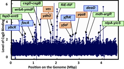
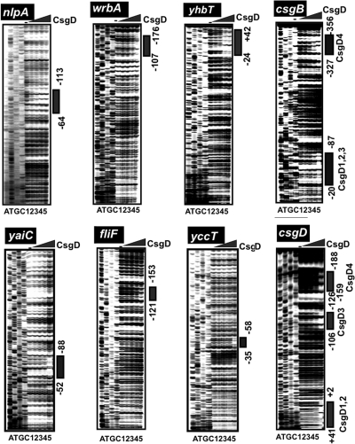
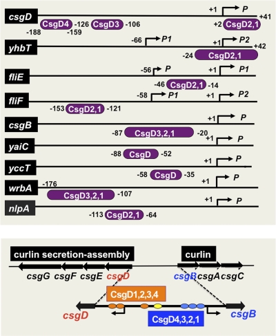
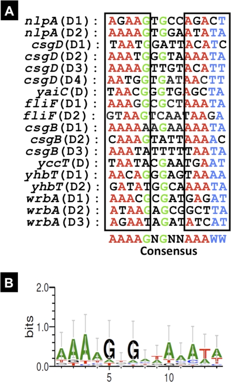
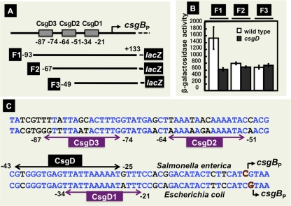
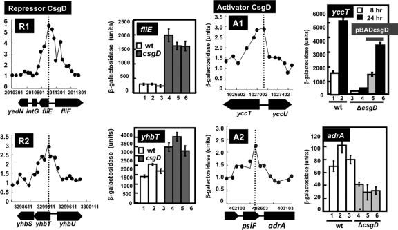
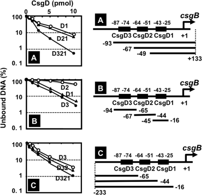
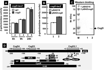
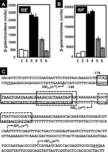
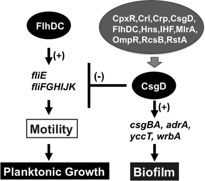
CsgD, the master regulator of biofilm formation, activates the synthesis of curli fimbriae and extracellular polysaccharides in Escherichia coli. To obtain insights into its regulatory role, we have identified a total of 20 novel regulation target genes on the E. coli genome by using chromatin immunoprecipitation (ChIP)-on-chip analysis with a high-density DNA microarray. By DNase I footprinting, the consensus CsgD-binding sequence predicted from a total of 18 target sites was found to include AAAAGNG(N(2))AAAWW. After a promoter-lacZ fusion assay, the CsgD targets were classified into two groups: group I genes, such as fliE and yhbT, are repressed by CsgD, while group II genes, including yccT and adrA, are activated by CsgD. The fliE and fliEFGH operons for flagellum formation are directly repressed by CsgD, while CsgD activates the adrA gene, which encodes an enzyme for synthesis of cyclic di-GMP, a bacterial second messenger, which in turn inhibits flagellum production and rotation. Taking these findings together, we propose that the cell motility for planktonic growth is repressed by CsgD, thereby promoting the switch to biofilm formation.


Figures

Fig. 1.
Fig. 1.
Maps of CsgD-binding sites on the E. coli genome. CsgD-associated sites on the genome were identified by using the ChIP-chip system (17, 18, 45). Peaks that were more than 2-fold higher than the background are indicated by arrows. For group 1 targets CsgD-binding sites are located within spacer regions, while CsgD-binding sites are located on open reading frames for group 2 targets (for classifications, see Table 2). Targets shown with blue and green backgrounds indicate group 1A and group 1B, respectively. On the other hand, group 2 targets are shown with an orange background.
Fig. 2.
Fig. 2.
DNase I footprinting of CsgD-binding sites. Fluorescently labeled DNA fragments of the indicated CsgD target promoters (1.0 pmol each) were incubated in the absence (lane 1) or presence of increasing concentrations of purified CsgD (lanes 2 to 5 contain 2.5, 5, 10, and 20 pmol) and then subjected to DNase I footprinting assays. Lanes A, T, G, and C represent the sequence ladders. Bold lines on the right indicate the CsgD-binding sequences as detected by their protection pattern after DNase I treatment. The nucleotide numbers indicate the distances from the respective transcription start sites. The locations of CsgD-binding sites are illustrated in Fig. 3, below.
Fig. 3.
Fig. 3.
Location of the CsgD-binding site(s) on the regulation target promoters. The locations of CsgD-binding sites on the promoters examined in the footprinting assay (Fig. 2) are shown along the respective promoter sequences. Transcription initiation sites have been determined for nlpA (6), csgD (22, 35), yaiC (10), fliE (33, 48), fliF (33, 48), csgB (2), yccT (data not shown), yhbT (29), and wrbA (29). Numbers on each line represent the distance (in bp) from the respective transcription start site. Note that the CsgD-binding site of fliE (−46 to −14) is the same as that of fliF (−121 to −153).
Fig. 4.
Fig. 4.
Consensus sequence of the CsgD box. CsgD-binding sites were identified in the wrbA, yhbT, yccT, csgB, fliF, yaiC, csgD and nlpA promoters, as shown in Fig. 2. The sequences of these promoter segments were analyzed to identify the consensus sequence of the CsgD box.
Fig. 5.
Fig. 5.
Analysis of functional roles of CsgD sites on the csgB promoter. (A) Three different segments (F1, F2, and F3) of the csgB promoter were inserted into pRS551 (47), and the resulting lacZ fusion plasmids were transformed into wild-type E. coli and csgD-defective mutant strains. (B) β-Galactosidase activities of the transformants. (C) Sequence of the E. coli csgB promoter and the locations of CsgD-binding sites are shown on the lower sequence, while the sequence for the Salmonella csgB promoter and the location of the CsgD-binding site (56) are shown in the upper sequence.
Fig. 6.
Fig. 6.
Classification of the regulation mode for CsgD. The promoter activity was measured in both the wild type and a csgD-defective mutant by using lacZ as a reporter. Based on the activity ratio between the wild type and csgD mutant, the test promoters were classified into repression mode (R1, fliE promoter; R2, yhbT promoter) and activation mode (A1, yccT promoter; A2, adrA promoter). For each promoter, both the ChIP-chip profile (left) and lacZ reporter activity (right) are shown. The sequences of peak regions are described in Fig. S1 of the supplemental material.
Fig. 7.
Fig. 7.
CsgD-binding activity of three CsgD boxes on the csgB promoter. Three sets of csgB promoter probes were constructed and examined for CsgD-binding activity by gel shift assay. (A) 5′ deletion set of the csgB promoter; (B) internal segments, each carrying one of three CsgD boxes; (C) 3′ deletion set of the csgB promoter. Construction of 5′ deletion (A) and 3′ deletion (C) probes was carried out by PCR using the primer pairs described in Materials and Methods, while the internal segment probes (B) were prepared by hybridization of complementary single-stranded DNA segments (see Table S1 for sequences).
Fig. 8.
Fig. 8.
Effects of CsgD on the csgD promoter. (A) E. coli BWWF1D (white bars) and BWcsgDF1D (gray bars) were grown in YESCA medium for 6 (lanes 1 and 2), 9 (lanes 3 and 4), or 24 h (lanes 7 and 8) for measurement of β-galactosidase activities. (B) E. coli BWcsgDF1D was transformed with either pBAD18 (lane 1) or pBADcsgD (lane 2) and grown in YESCA medium at 28°C for 12 h, and then β-galactosidase activities were measured. (C) Confirmation of CsgD expression in pBADcsgD-transformed E. coli BWcsgDF1D by Western blot analysis. (D) Locations of CsgD-binding sites along the csgD promoter. The binding sites of other transcription factors on the cagD promoter have been reported previously (36, 37).
Fig. 9.
Fig. 9.
Effects of CsgD on the fliE and fliF promoters. (A) E. coli BWWfliE (lanes 1 and 2) and BWcsgDfliE (lanes 3 to 6) strains were transformed with either pBAD18 (lanes 1 to 4) or pBADcsgD (lanes 5 and 6), grown in YESCA medium at 28°C for 8 h (lanes 1, 3, and 5) or 24 h (lanes 2, 4, and 6), and β-galactosidase activities were determined. (B) E. coli BWWfliF (lanes 1 and 2) and BWcsgDfliF (lanes 3 to 6) strains were transformed with either pBAD18 (lanes 1 to 4) or pBADcsgD (lanes 5 and 6), grown in YESCA medium at 28°C for 8 h (lanes 1, 3, and 5) or 24 h (lanes 2, 4, and 6), and β-galactosidase activity was assayed. (C) Locations of CsgD-binding sites within the spacer region between fliE and fliF. The CsgD-binding site of CsgD is indicated by the solid-lined box, while two binding sites of FlhDC, the activator for the fliE and fliF operons, are indicated by the boxes with dotted lines. The fliE and fliF operons are both transcribed by both σ70 and σ28 RNA polymerases.
Fig. 10.
Fig. 10.
The regulatory roles of CsgD in biofilm formation and cell motility. The master regulator CsgD for biofilm formation represses the genes for flagellum formation and motility, thereby switching the bacterium's lifestyle to the biofilm mode.

References

    1. Armitage J. P., Berry R. M. 2010. Time for bacteria to slow down. Cell 141:24–26 - PubMed
    1. Arnqvist A., Olsen A., Normark S. 1994. Sigma S-dependent growth-phase induction of the csgBA promoer in Escherichia coli can be achieved in vivo by sigma 70 in the absence of the nucleoid-associated protein H-NS. Mol. Microbiol. 13:1021–1032 - PubMed
    1. Baba T., et al. 2006. Construction of Escherichia coli K-12 in-frame, single-gene knockout mutants: the Keio collection. Mol. Syst. Biol. 2:2006.0008 - PMC - PubMed
    1. Barnhart M. M., Chapman M. R. 2006. Curli biogenesis and function. Annu. Rev. Microbiol. 60:131–147 - PMC - PubMed
    1. Beloin C., et al. 2004. Global impact of mature biofilm lifestyle on Escherichia coli K-12 gene expression. Mol. Microbiol. 51:659–674 - PubMed

Publication types

MeSH terms

LinkOut - more resources