Comparative Study
. 2011 Jun;4(3):388-96.
doi: 10.1161/CIRCEP.110.959650. Epub 2011 Mar 18.

Autonomic remodeling in the left atrium and pulmonary veins in heart failure: creation of a dynamic substrate for atrial fibrillation

Affiliations
Comparative Study

Autonomic remodeling in the left atrium and pulmonary veins in heart failure: creation of a dynamic substrate for atrial fibrillation

Jason Ng et al. Circ Arrhythm Electrophysiol. 2011 Jun.

Abstract

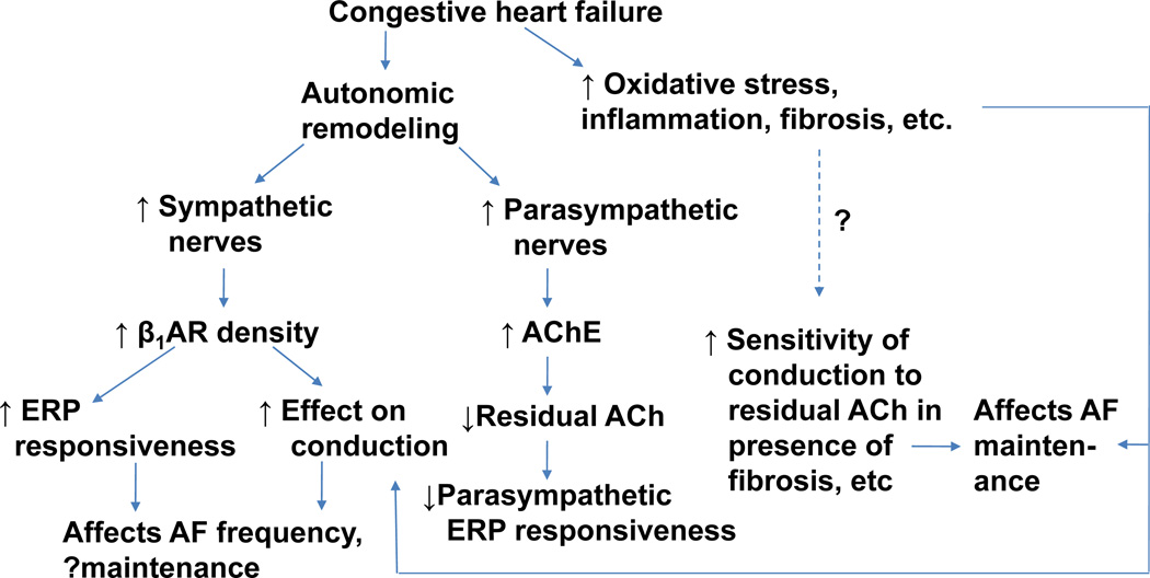
Background: Atrial fibrillation (AF) is commonly associated with congestive heart failure (CHF). The autonomic nervous system is involved in the pathogenesis of both AF and CHF. We examined the role of autonomic remodeling in contributing to AF substrate in CHF.

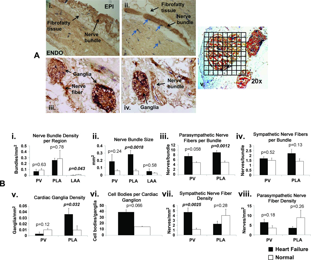
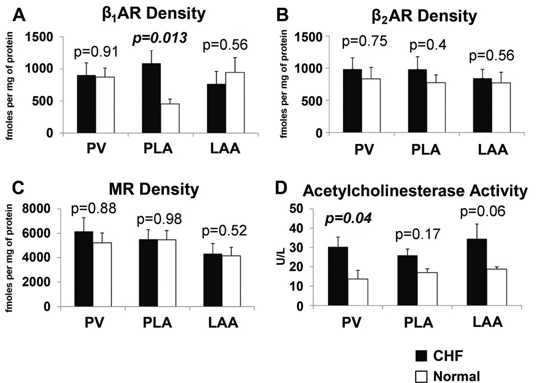
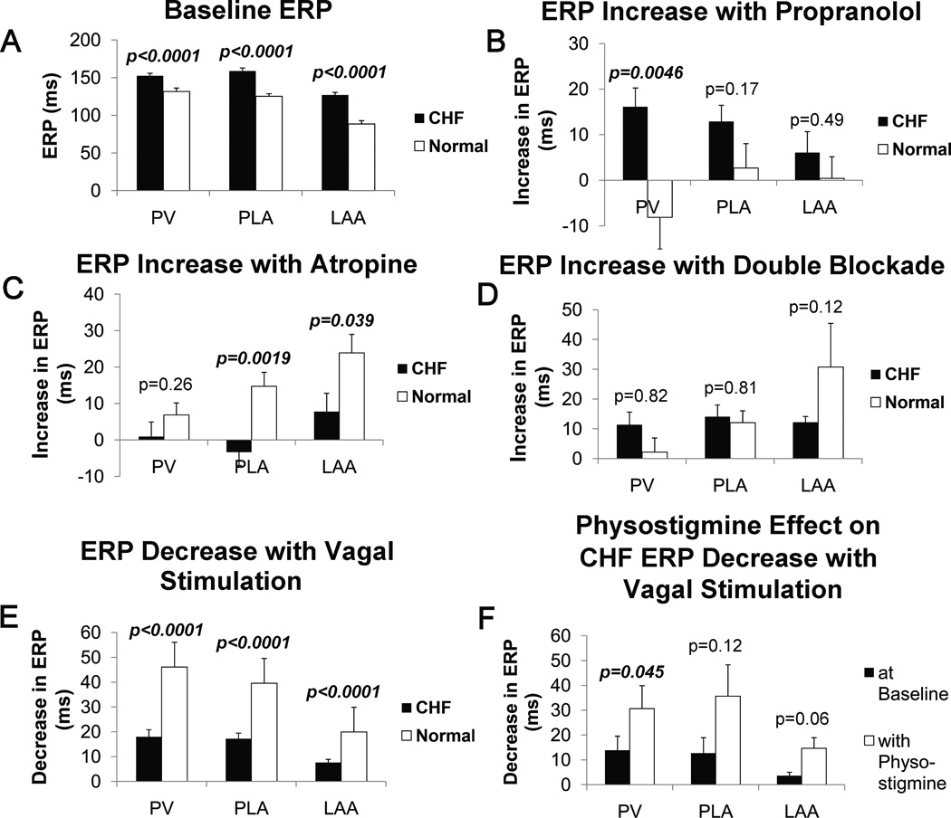
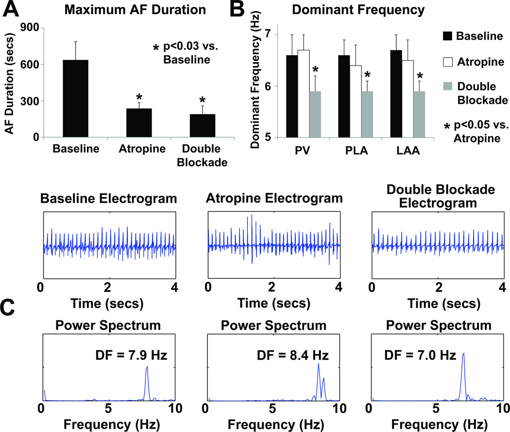
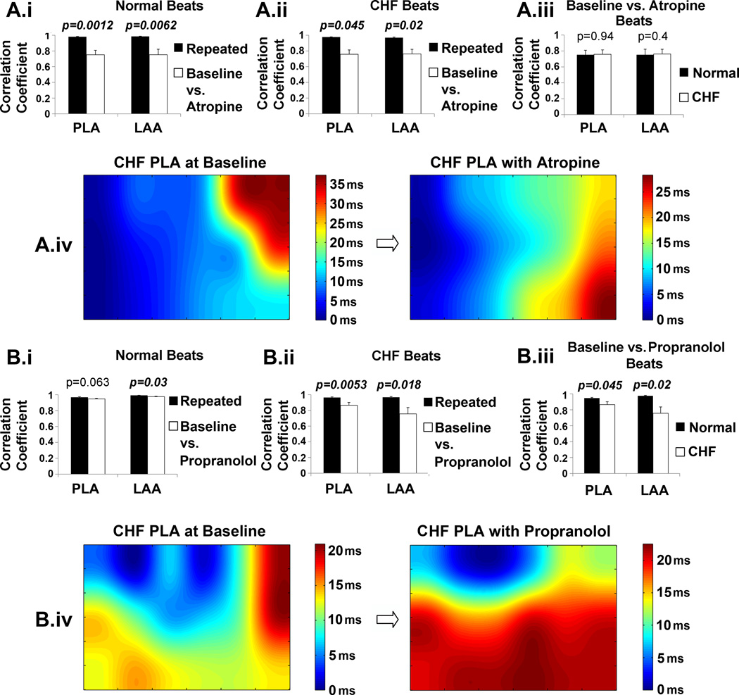
Methods and results: Electrophysiological mapping was performed in the pulmonary veins and left atrium in 38 rapid ventricular-paced dogs (CHF group) and 39 control dogs under the following conditions: vagal stimulation, isoproterenol infusion, β-adrenergic blockade, acetylcholinesterase (AChE) inhibition (physostigmine), parasympathetic blockade, and double autonomic blockade. Explanted atria were examined for nerve density/distribution, muscarinic receptor and β-adrenergic receptor densities, and AChE activity. In CHF dogs, there was an increase in nerve bundle size, parasympathetic fibers/bundle, and density of sympathetic fibrils and cardiac ganglia, all preferentially in the posterior left atrium/pulmonary veins. Sympathetic hyperinnervation was accompanied by increases in β(1)-adrenergic receptor R density and in sympathetic effect on effective refractory periods and activation direction. β-Adrenergic blockade slowed AF dominant frequency. Parasympathetic remodeling was more complex, resulting in increased AChE activity, unchanged muscarinic receptor density, unchanged parasympathetic effect on activation direction and decreased effect of vagal stimulation on effective refractory period (restored by AChE inhibition). Parasympathetic blockade markedly decreased AF duration.

Conclusions: In this heart failure model, autonomic and electrophysiological remodeling occurs, involving the posterior left atrium and pulmonary veins. Despite synaptic compensation, parasympathetic hyperinnervation contributes significantly to AF maintenance. Parasympathetic and/or sympathetic signaling may be possible therapeutic targets for AF in CHF.


Conflict of interest statement

Conflict of Interest Disclosures: None

Figures

Figure 1
Figure 1
Panel A: Examples of sympathetic and parasympathetic nerve staining. A.i. – example of a nerve bundle located in the fibrofatty tissue overlying the epicardium (EPI) (10×). ENDO – endocardium. A.ii. – example of nerve bundles located in fibrofatty tissue on the epicardial aspect of PV (4×). Sympathetic fibers are in blue (arrows). A.iii. – examples of cardiac ganglia, with parasympathetic fibers arising from cardiac ganglion on the left side (20×). A.iv. - example of cardiac ganglia on the left and nerve bundle on the right; nerve fibers showing co-localized sympathetic (blue) and parasympathetic fibers (brown) (20×). A.v. - illustration of the use of 1 mm × 1 mm grids to quantify the cross-sectional area of a nerve bundle at 20× magnification. Panel B: Quantitative analysis of nerve staining. B.i. – nerve bundle density, B.ii. – nerve bundle size, B.iii. – number of parasympathetic nerve fibers/bundle, B.iv. – number of sympathetic nerve fibers/bundle, B.v. – density of cardiac ganglia, B.vi. – number of cell bodies/cardiac ganglion, B.vii - density of sympathetic fibers, B.viii. – density of parasympathetic fibers.
Figure 2
Figure 2
Comparison of beta-adrenergic receptors (βAR), muscarinic receptors (MR), and acetylcholinesterase (AChE) in the PV, PLA and LAA for CHF and normal dogs.
Figure 3
Figure 3
Result of ERP testing obtained from the PVs, PLA, and LAA for CHF and normal dogs. A - ERPs at baseline; B – increase in ERP with propranolol; C – increase in ERP with atropine; D – increase in ERP with double blockade (combined propranolol and atropine); E – decrease in ERP with vagal stimulation effect; F – decrease in ERP with vagal stimulation effect for CHF dogs with and without physostigmine.
Figure 4
Figure 4
Results of AF analysis in CHF dogs. A – Maximum AF durations obtained by burst pacing at baseline, with atropine, and with double blockade, B – Average dominant frequencies (DF) in the PVs, PLA and LAA at baseline, with atropine, and with double blockade. C – examples of PLA electrograms and corresponding power spectrum during baseline, with atropine, and with double blockade.
Figure 5
Figure 5
Effect of autonomic blockade on activation patterns in the PLA and LAA for CHF and normal dogs. The corresponding results for atropine and propranolol are shown in panels A and B, respectively. A.i and B.i show the effect of autonomic blockade on conduction in the normal dog by comparing correlation coefficients of activation times from the repeated beats compared with the correlation coefficients obtained between baseline vs. autonomic blockade beats, A.ii and B.ii show the effect of autonomic blockade on conduction in the CHF dogs. A.iii and B.iii compare the correlation coeffients of activation times of the normal dogs vs. the CHF dogs. A.iv and B.iv show examples of activation maps from the PLA before and after autonomic blockade in a CHF dog.
Figure 6
Figure 6
Proposed model of creation of autonomic substrate for AF in CHF. The model suggests the likely presence of synergistic interactions between structural changes (fibrosis) and autonomic remodeling in the creation of AF substrate in heart failure. ACh = acetylcholine, AChE = acetylcholinesterase, β1AR – beta-1 adrenergic receptor, ERP = effective refractory period

References

    1. Ehrlich JR, Nattel S, Hohnloser SH. Atrial fibrillation and congestive heart failure: specific considerations at the intersection of two common and important cardiac disease sets. J Cardiovasc Electrophysiol. 2002;13:399–405. - PubMed
    1. Wang TJ, Larson MG, Levy D, Vasan RS, Leip EP, Wolf PA, D'Agostino RB, Murabito JM, Kannel WB, Benjamin EJ. Temporal relations of atrial fibrillation and congestive heart failure and their joint influence on mortality: the Framingham Heart Study. Circulation. 2003;107:2920–2925. - PubMed
    1. Everett THt, Olgin JE. Atrial fibrosis and the mechanisms of atrial fibrillation. Heart Rhythm. 2007;4:S24–S27. - PMC - PubMed
    1. Li D, Melnyk P, Feng J, Wang Z, Petrecca K, Shrier A, Nattel S. Effects of experimental heart failure on atrial cellular and ionic electrophysiology. Circulation. 2000;101:2631–2638. - PubMed
    1. Li D, Fareh S, Leung TK, Nattel S. Promotion of atrial fibrillation by heart failure in dogs: atrial remodeling of a different sort. Circulation. 1999;100:87–95. - PubMed

Publication types

MeSH terms