

Entrapment of conjunctival goblet cells by desiccation-induced cornification
- PMID: 21421863
- PMCID: PMC3109039
- DOI: 10.1167/iovs.10-5782
Entrapment of conjunctival goblet cells by desiccation-induced cornification
Abstract
Purpose: To evaluate the effects of desiccating stress on conjunctival goblet cell density and morphology and the expression of cornified envelope precursors by the ocular surface epithelia.
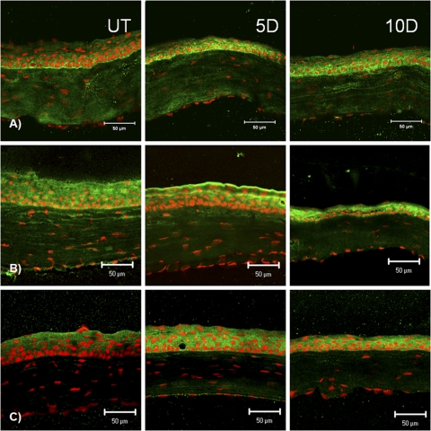
Methods: Experimental dry eye (EDE) was created in C57BL/6 mice. Real-time PCR evaluated the expression of cornified envelope (CE) precursor proteins (involucrin and small proline-rich [Sprr] -1a, -1b, -2a, -2b, -2f, and -2g proteins), the cross-linking transglutaminase 1 enzyme (Tg-1) and Muc5AC mRNA transcripts by the ocular surface epithelia. Laser scanning confocal microscopy evaluated the expression of the CE precursor proteins Tg-1 and Muc5AC in cryosections. Tg-1 activity was measured by a fluorescein cadaverine assay. Muc5AC concentration was measured by ELISA.
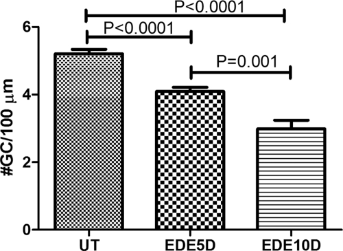
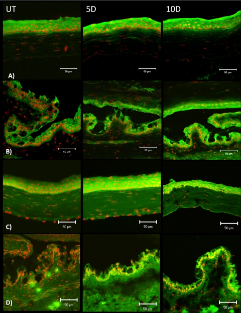
Results: Levels of involucrin; Sprr-1a, -1b, -2a, -2b, -2f, and -2g; and Tg1-1 mRNA transcripts in ocular surface tissues increased in response to desiccating stress. Expression and activity of Tg in the conjunctiva markedly increased after EDE. Desiccating stress caused progressive loss of mucin-filled goblet cells. The apical portion of the remaining conjunctival goblet cells became entrapped by adjacent stratified apical epithelia expressing increased levels of cornified envelope precursors.
Conclusions: Exposure to desiccating stress stimulates ocular surface epithelia to produce cornified envelope precursors and the tissue transglutaminase enzyme that cross-links them. This effect is accompanied by loss of mucin-filled goblet cells and entrapment of mucin contents in the remaining ones by cornifying cells that block the egress of mucin contents to the ocular surface. This mechanism may contribute to the conjunctival mucin deficiency that develops in dry eye.
Figures
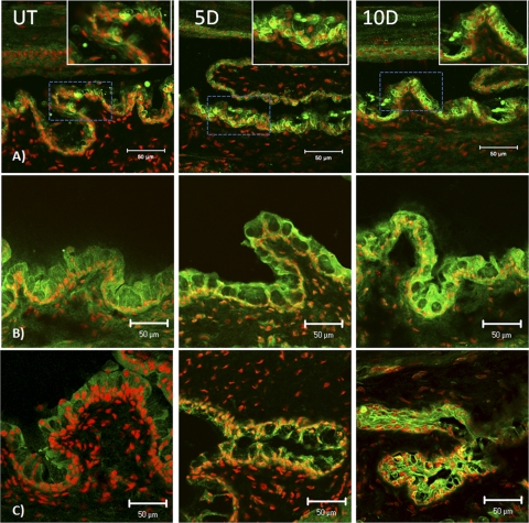
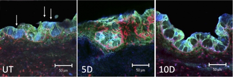
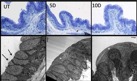
 ). Secretory granules in these goblet cells were reduced and varied greatly in electron density compared with those in UT control eyes.
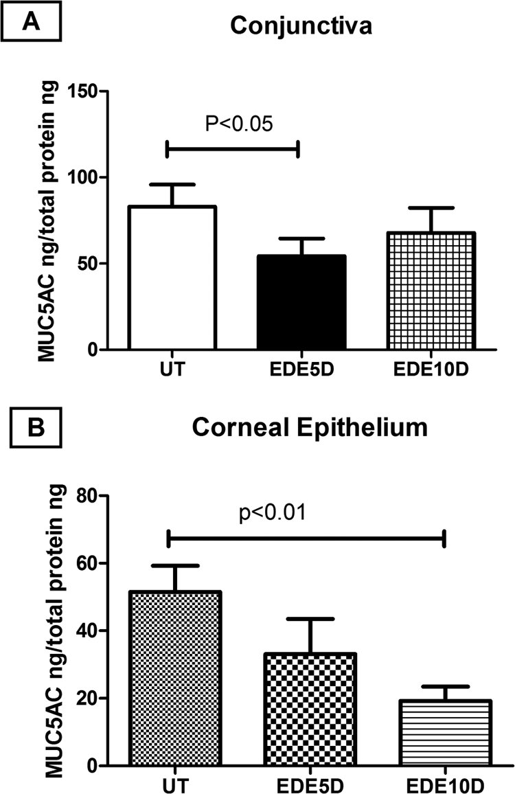
References
-
- Steven AC, Steinert PM. Protein composition of cornified cell envelopes of epidermal keratinocytes. J Cell Sci. 1994;107:693–700 - PubMed
-
- Castro-Munozledo F. Development of a spontaneous permanent cell line of rabbit corneal epithelial cells that undergoes sequential stages of differentiation in cell culture. J Cell Sci. 1994;107:2343–2351 - PubMed
-
- Adhikary G, Crish J, Lass J, Eckert RL. Regulation of involucrin expression in normal human corneal epithelial cells: a role for activator protein one. Invest Ophthalmol Vis Sci. 2004;45:1080–1087 - PubMed
Publication types
MeSH terms
Substances
Grants and funding
LinkOut - more resources
Full Text Sources
Miscellaneous
