

Blocking the leukotriene B4 receptor 1 inhibits late-phase airway responses in established disease
- PMID: 21421908
- PMCID: PMC3208610
- DOI: 10.1165/rcmb.2010-0455OC
Blocking the leukotriene B4 receptor 1 inhibits late-phase airway responses in established disease
Abstract
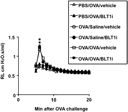
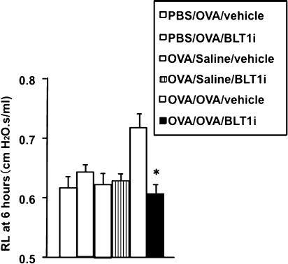
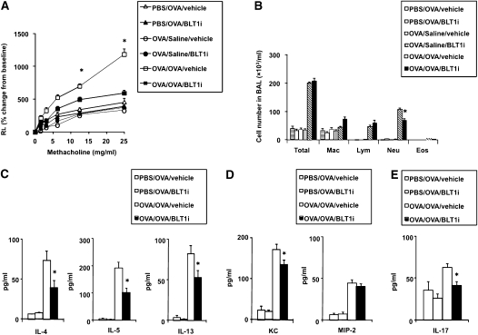
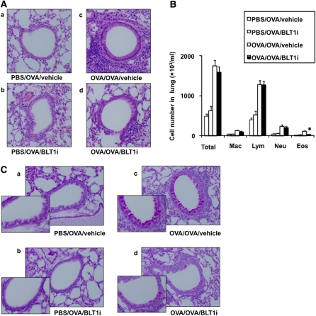
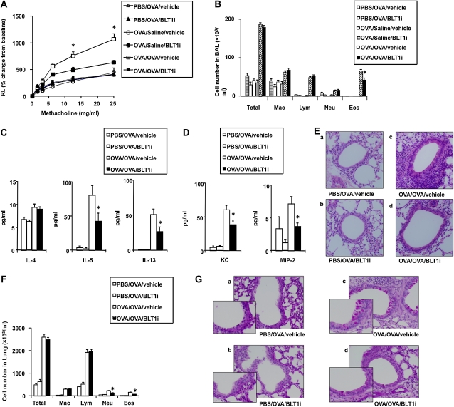
Most of the studies investigating the effectiveness of blocking the leukotriene B4 (LTB4) receptor 1 (BLT1) have been performed in models of primary or acute allergen challenge. The role of the LTB4-BLT1 pathway in secondary challenge models, where airway hyperresponsiveness (AHR) and airway inflammation have been established, has not been defined. We investigated the effects of blocking BLT1 on early- and late-phase development of AHR and airway inflammation in previously sensitized and challenged mice. Female BALB/c mice were sensitized (Days 1 and 14) and challenged (primary, Days 28-30) with ovalbumin. On Day 72, mice were challenged (secondary) with a single OVA aerosol, and the early and late phases of AHR and inflammation were determined. Specific blockade of BLT1 was attained by oral administration of a BLT1 antagonist on Days 70 through 72. Administration of the antagonist inhibited the secondary ovalbumin challenge-induced alterations in airway responses during the late phase but not during the early phase, as demonstrated by decreases in AHR and in bronchoalveolar lavage neutrophilia and eosinophilia 6 and 48 hours after secondary challenge. The latter was associated with decreased levels of KC protein, macrophage inflammatory protein 2, and IL-17 in the airways. These data identify the importance of the LTB4-BLT1 pathway in the development of late-phase, allergen-induced airway responsiveness after secondary airway challenge in mice with established airway disease.
Figures
Similar articles
-
Requirement for leukotriene B4 receptor 1 in allergen-induced airway hyperresponsiveness.Am J Respir Crit Care Med. 2005 Jul 15;172(2):161-7. doi: 10.1164/rccm.200502-205OC. Epub 2005 Apr 22. Am J Respir Crit Care Med. 2005. PMID: 15849325 Free PMC article.
-
Anti-Asthma Simplified Herbal Medicine Intervention-induced long-lasting tolerance to allergen exposure in an asthma model is interferon-γ, but not transforming growth factor-β dependent.Clin Exp Allergy. 2010 Nov;40(11):1678-88. doi: 10.1111/j.1365-2222.2010.03545.x. Clin Exp Allergy. 2010. PMID: 20573156 Free PMC article.
-
Intracerebroventricular injection of leukotriene B4 attenuates antigen-induced asthmatic response via BLT1 receptor stimulating HPA-axis in sensitized rats.Respir Res. 2010 Apr 20;11(1):39. doi: 10.1186/1465-9921-11-39. Respir Res. 2010. PMID: 20403205 Free PMC article.
-
CD8+ T lymphocytes and leukotriene B4: novel interactions in the persistence and progression of asthma.J Allergy Clin Immunol. 2006 Mar;117(3):577-82. doi: 10.1016/j.jaci.2005.12.1340. J Allergy Clin Immunol. 2006. PMID: 16522456 Review.
-
Monitoring asthma in childhood: lung function, bronchial responsiveness and inflammation.Eur Respir Rev. 2015 Jun;24(136):204-15. doi: 10.1183/16000617.00003914. Eur Respir Rev. 2015. PMID: 26028633 Free PMC article. Review.
Cited by
-
Inhibition of neutrophil elastase attenuates airway hyperresponsiveness and inflammation in a mouse model of secondary allergen challenge: neutrophil elastase inhibition attenuates allergic airway responses.Respir Res. 2013 Jan 24;14(1):8. doi: 10.1186/1465-9921-14-8. Respir Res. 2013. PMID: 23347423 Free PMC article.
-
Lower myostatin and higher MUC1 levels are associated with better response to mepolizumab and omalizumab in asthma: a protein-protein interaction analyses.Respir Res. 2023 Dec 6;24(1):305. doi: 10.1186/s12931-023-02620-1. Respir Res. 2023. PMID: 38057814 Free PMC article.
-
Assessment of murine lung mechanics outcome measures: alignment with those made in asthmatics.Front Physiol. 2013 Feb 12;3:491. doi: 10.3389/fphys.2012.00491. eCollection 2012. Front Physiol. 2013. PMID: 23408785 Free PMC article.
-
Cigarette smoke-induced lung inflammation in COPD mediated via LTB4/BLT1/SOCS1 pathway.Int J Chron Obstruct Pulmon Dis. 2015 Dec 22;11:31-41. doi: 10.2147/COPD.S96412. eCollection 2016. Int J Chron Obstruct Pulmon Dis. 2015. PMID: 26730186 Free PMC article.
-
An extracellular matrix fragment drives epithelial remodeling and airway hyperresponsiveness.Sci Transl Med. 2018 Aug 22;10(455):eaaq0693. doi: 10.1126/scitranslmed.aaq0693. Sci Transl Med. 2018. PMID: 30135247 Free PMC article.
References
-
- Busse WW, Lemanske RF., Jr Asthma. N Engl J Med 2001;344:350–362 - PubMed
-
- Lee NA, Gelfand EW, Lee JJ. Pulmonary T cells and eosinophils: coconspirators or independent triggers of allergic respiratory pathology? J Allergy Clin Immunol 2001;107:945–957 - PubMed
-
- De Sanctis GT, Itoh A, Green FH, Qin S, Kimura T, Grobholz JK, Martin TR, Maki T, Drazen JM. T-lymphocytes regulate genetically determined airway hyperresponsiveness in mice. Nat Med 1997;3:460–462 - PubMed
-
- Robinson DS, Hamid QA, Ying S, Tsicopoulos A, Barkans J, Bentley AM, Corrigan C, Durham SR, Kay AB. Predominant TH2-like bronchoalveolar T-lymphocyte population in atopic asthma. N Engl J Med 1992;326:298–304. - PubMed
-
- van Rensen EL, Sont JK, Evertse CE, Willems LN, Mauad T, Hiemstra PS. Sterk PJ, and the AMPUL Study Group. Bronchial CD8 cell infiltrate and lung function decline in asthma. Am J Respir Crit Care Med 2005;172:837–841 - PubMed
