Review
. 2011 Jan 1;3(1):a006833.
doi: 10.1101/cshperspect.a006833.

Prions

Affiliations
Review

Prions

David W Colby et al. Cold Spring Harb Perspect Biol. .

Abstract

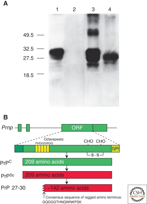
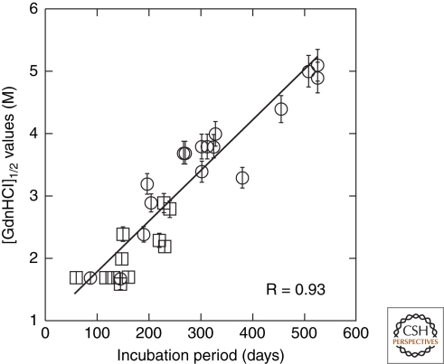
The discovery of infectious proteins, denoted prions, was unexpected. After much debate over the chemical basis of heredity, resolution of this issue began with the discovery that DNA, not protein, from pneumococcus was capable of genetically transforming bacteria (Avery et al. 1944). Four decades later, the discovery that a protein could mimic viral and bacterial pathogens with respect to the transmission of some nervous system diseases (Prusiner 1982) met with great resistance. Overwhelming evidence now shows that Creutzfeldt-Jakob disease (CJD) and related disorders are caused by prions. The prion diseases are characterized by neurodegeneration and lethality. In mammals, prions reproduce by recruiting the normal, cellular isoform of the prion protein (PrP(C)) and stimulating its conversion into the disease-causing isoform (PrP(Sc)). PrP(C) and PrP(Sc) have distinct conformations: PrP(C) is rich in α-helical content and has little β-sheet structure, whereas PrP(Sc) has less α-helical content and is rich in β-sheet structure (Pan et al. 1993). The conformational conversion of PrP(C) to PrP(Sc) is the fundamental event underlying prion diseases. In this article, we provide an introduction to prions and the diseases they cause.


Figures

Figure 1.
Figure 1.
Prion protein isoforms. (A) Western immunoblot of brain homogenates from uninfected (lanes 1 and 2) and prion-infected (lanes 3 and 4) Syrian hamsters. Samples in lanes 2 and 4 were digested with 50 µg/µl proteinase K for 30 min at 37°C, completely hydrolyzing PrPC. Proteinase digestion cleaves ∼67 amino acids from the amino terminus of PrPSc to generate PrP 27–30 (lane 4). Blot developed with anti-PrP polyclonal antiserum R073 (Serban et al. 1990). (B) Bar diagrams of the hamster Prnp gene and PrP isoforms. The Prnp ORF encodes a protein of 254 residues, which is shortened to 209 residues during posttranslational processing. PrPSc is an alternate conformation of PrPC with identical primary structure. Limited proteolysis of PrPSc cleaves the amino terminus and produces PrP 27-30, composed of approximately 142 residues. Panel A, reprinted with permission, from Prusiner 2004.
Figure 2.
Figure 2.
Variation of in the prion protein gene. (A) Species variations of the prion protein gene. The x-axis represents the human PrP sequence, with the five octarepeats and H1–H4 regions of the putative secondary structure shown, as well as the three α-helices A, B, and C and the two β-strands S1 and S2 as determined by NMR. Vertical bars above the axis indicate the number of species that differ from the human sequence at each position. Below the axis, the length of the bars indicates the number of alternative amino acids at each position in the alignment. (B) PrP mutations causing inherited human prion disease (above the line) and PrP polymorphisms (below the line) found in humans, mice, sheep, elk, and cattle. Residue numbers in parentheses correspond to the human codons. Data in Panel A compiled by P. Bamborough and F.E. Cohen and reprinted, with permission, from Prusiner 2004.
Figure 3.
Figure 3.
Bioluminescence in Tg(Gfap-luc) mice inoculated intracerebrally with RML prions (n = 12) indicated a reactive astrocytic gliosis. (A) Bioluminescence measured from the brains of prion-inoculated mice (black circles) began to increase at 55 d postinoculation (dpi). Bioluminescence in control Tg(Gfap-luc) mice inoculated with 1% normal brain homogenate (NBH) (n = 4, gray squares) remained low throughout the incubation period. (B–D) Photos of representative Tg(Gfap-luc) mice, with overlays of the circular area above the brain from which bioluminescence was quantified. Bioluminescence measured, ×106 photons/s, from each mouse brain is shown below each image. The bioluminescence measured from the brains of prion-infected mice significantly increased (**, P < 0.001, Bonferroni t test) from 48 dpi (B) to 55 dpi (C). Similarly, bioluminescence measured from infected mice at 55 dpi (C) was also significantly (*, P < 0.005) greater than in control mice inoculated with NBH and imaged at 56 dpi (D). No significant difference (N.S., P < 0.5) was measured between RML-inoculated mice at 48 dpi (B) and control mice at 56 dpi (D). Based on this result, astrocytic gliosis was detectable at bioluminescence measurements >2.0 × 10⁶ photons/s. Reprinted, with permission, from Tamgüney et al. 2009a.
Figure 4.
Figure 4.
Structures of PrPC. (A) NMR structure of Syrian hamster (SHa) recombinant (rec) PrP(90–231), which presumably resembles PrPC. Blue, α-helices; yellow, loops; green, β-strands (James et al. 1997). (B) Schematic diagram showing degree of structure for entire PrP polypeptide chain based on {1H}-15N NOE data. Red, most flexible regions of the protein; blue, least flexible regions (James et al. 1997). Arbitrary structure is shown for residues 23–89. Reprinted, with permission, from Prusiner 2004.
Figure 5.
Figure 5.
Structural models of PrPSc. (A) Residues 89–174 of PrP threaded into a left-handed β-helix based on UDP N-acetylglucosamine O-acyltransferase from Escherichia coli (PDB ID code 1LXA). (B) Model of the monomer of PrP 27–30 with the α-helical region (residues 177–227) as determined by NMR spectroscopy shown in red. (C) The crystal structure of the trimeric carbonic anhydrase from Methanosarcina thermophila. (D) Trimeric model of PrP 27–30 built by superimposing three monomeric models onto the structure shown in C. (E) Projection map of PrP 27–30 obtained by processing and averaging three independent 2D crystals of PrP 27–30. (F) Statistically significant differences between PrP 27–30 and PrPSc106 overlaid onto the projection map of PrP 27–30. The differences attributed to the internal deletion of PrPSc106 (residues 141–176) are shown in red; the differences in glycosylation between PrP 27–30 and PrPSc106 are shown in blue. (G) Superimposition of the trimeric left-handed model onto the EM maps. The trimeric left-handed α-helical model of PrP 27–30 is superimposed on a 1:1 scale with the electron crystallographic maps of PrP 27–30. (H) The scaled trimeric model was copied onto the neighboring units of the crystals to show the crystallographic packing suggested by the model. Bars in panels E–H represent 50 Å. Reprinted with permission, from Govaerts et al. 2004a.
Figure 6.
Figure 6.
The conformational stability of prions is directly proportional to the length of the incubation time in mice. The [GdnHCl]1/2 values for prions were plotted as a function of the incubation times. Synthetic prions (circles) in the brains of Tg9949, Tg4053, and non-Tg FVB mice were plotted with many naturally occurring prions passaged (squares) in both non-Tg and Tg mice. R = 0.93. Reprinted, with permission, from Legname et al. 2006.

Similar articles

Cited by

References

    1. Alpers MP 1968. Kuru: implications of its transmissibility for the interpretation of its changing epidemiological pattern. In The central nervous system: some experimental models of neurological diseases. (ed. Bailey OT, Smith DE), pp. 234–251 Williams and Wilkins Company, Baltimore
    1. Anderson RM, Donnelly CA, Ferguson NM, Woolhouse MEJ, Watt CJ, Udy HJ, MaWhinney S, Dunstan SP, Southwood TRE, Wilesmith JW, et al.1996. Transmission dynamics and epidemiology of BSE in British cattle. Nature 382: 779–788 - PubMed
    1. Antonyuk SV, Trevitt CR, Strange RW, Jackson GS, Sangar D, Batchelor M, Cooper S, Fraser C, Jones S, Georgiou T, et al.2009. Crystal structure of human prion protein bound to a therapeutic antibody. Proc Natl Acad Sci 106: 2554–2558 - PMC - PubMed
    1. Avery OT, MacLeod CM, McCarty M 1944. Studies on the chemical nature of the substance inducing transformation of pneumococcal types. Induction of transformation by a deoxyribonucleic acid fraction isolated from pneumococcus type III. J Exp Med 79: 137–158 - PMC - PubMed
    1. Barria MA, Mukherjee A, Gonzalez-Romero D, Morales R, Soto C 2009. De novo generation of infectious prions in vitro produces a new disease phenotype. PLoS Pathog 5: e1000421. - PMC - PubMed