. 2011 Jan 1;3(1):a002261.
doi: 10.1101/cshperspect.a002261.

The coordination of T-cell function by serine/threonine kinases

Affiliations

The coordination of T-cell function by serine/threonine kinases

David Finlay et al. Cold Spring Harb Perspect Biol. .

Abstract

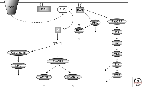
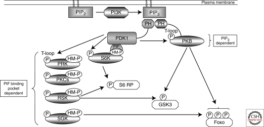
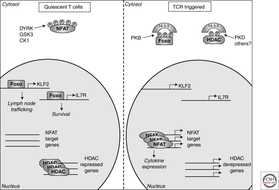
The function of T-lymphocytes during adaptive immune responses is directed by antigen receptors, costimulatory molecules, and cytokines. These extrinsic stimuli are coupled to a network of serine/threonine kinases that control the epigenetic, transcriptional, and metabolic programs that determine T-cell function. It is increasingly recognized that serine/threonine kinases, notably those that are controlled by lipid second messengers such as polyunsaturated diacylglycerols (DAG) and phosphatidylinositol-(3,4,5)-trisphosphate (PIP(3)), are at the core of T-cell signal transduction. In the present review the object will be to discuss some important examples of how pathways of serine/threonine phosphorylation control molecular functions of proteins and control protein localization to coordinate T-cell function in adaptive immune responses.


Figures

Figure 1.
Figure 1.
TCR activation of Ca2+/DAG regulated kinases. T-cell receptor (TCR) ligation activates phopholipase Cγ (PLCγ), which cleaves phosphotidylinositol-(3,4)-bisphosphate (PIP2) generating two key second messenger molecules, diacylglycerol (DAG) and inositol-(1,4,5)-trisphosphate (IP3). IP3 releases intracellular Ca2+ stores into the cytosol activating; the Ca2+ dependent phosphatase calcineurin inducing the expression of NFAT target genes, and the Ca2+ dependent kinase CaMKK which phosphorylates and activates AMPK and CaMK IV. The accumulation of DAG in the plasma membrane mediates the activation PKC and PKD protein kinases. DAG is also required for the activation of the Erk1/2 and RSK through activation of the Ras GTPase.
Figure 2.
Figure 2.
PDK1 signaling. PDK1 acts as a “master kinase” activating multiple AGC kinases through phosphorylation of a conserved Threonine residue (“T-loop” site) within the catalytic domain. PDK1 activation of PKB requires the second messenger molecule PIP3 that accumulates in the plasma membrane because of the action of the lipid kinase PI3K. PDK1 activation of other AGC kinases relies on prior phosphorylation of a Ser/Thr residue within a hydrophobic motif (HM). This phosphorylated motif creates a docking site for PDK1 to bind through its PIF binding pocket, thus allowing PDK1-mediated phosphorylation of the T-loop site and kinase activation. Significant redundancy exists in PDK1 signaling as many downstream substrates, such as S6 ribosomal protein, GSK3, and FoxO transcription factors, can be phosphorylated by multiple PDK1-regulated AGC kinases.
Figure 3.
Figure 3.
LKB1 is a “master kinase.” LKB1 phosphorylates and activates 13 kinases within the AMPK family of protein kinases.
Figure 4.
Figure 4.
Phosphorylation determined intracellular localization. In quiescent T cells constitutively active kinases (DYRK, GSK3, and CK1) maintain NFAT transcription factors in a phosphorylated state excluding them from the nucleus. In contrast, the kinases that regulate FoxO transcription factors and histone deacetylases (HDACs) are inactive in quiescent cells allowing FoxOs to access the nucleus and control genes involved in T-cell trafficking and survival whereas HDACs repress gene expression through modulating chromatin structure. Following triggering of the T-cell receptor (TCR) FoxO transcription factors and HDACs are phosphorylated, resulting in their cytosolic sequestration by 14-3-3 proteins, thus preventing their nuclear actions. Meanwhile, the Ca2+ activated phosphatase, calcineurin, dephosphorylates NFAT transcription factors allowing nuclear entry and the expression of key cytokine genes.

References

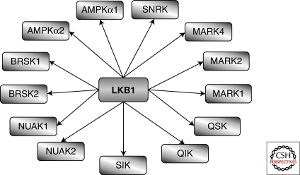
    1. Alessi DR, Sakamoto K, Bayascas JR 2006. LKB1-dependent signaling pathways. Annu Rev Biochem 75: 137–163 - PubMed
    1. Arbones ML, Ord DC, Ley K, Ratech H, Maynard-Curry C, Otten G, Capon DJ, Tedder TF 1994. Lymphocyte homing and leukocyte rolling and migration are impaired in L-selectin-deficient mice. Immunity 1: 247–260 - PubMed
    1. Astoul E, Laurence AD, Totty N, Beer S, Alexander DR, Cantrell DA 2003. Approaches to define antigen receptor-induced serine kinase signal transduction pathways. J Biol Chem 278: 9267–9275 - PubMed
    1. Bayascas JR, Wullschleger S, Sakamoto K, Garcia-Martinez JM, Clacher C, Komander D, van Aalten DM, Boini KM, Lang F, Lipina C, et al.2008. Mutation of the PDK1 PH domain inhibits protein kinase B/Akt, leading to small size and insulin resistance. Mol Cell Biol 28: 3258–3272 - PMC - PubMed
    1. Brunet A, Park J, Tran H, Hu LS, Hemmings BA, Greenberg ME 2001. Protein kinase SGK mediates survival signals by phosphorylating the forkhead transcription factor FKHRL1 (FOXO3a). Mol Cell Biol 21: 952–965 - PMC - PubMed

MeSH terms

Substances

LinkOut - more resources