Review
. 2011 Feb 1;3(2):a004911.
doi: 10.1101/cshperspect.a004911.

Basement membranes: cell scaffoldings and signaling platforms

Affiliations
Review

Basement membranes: cell scaffoldings and signaling platforms

Peter D Yurchenco. Cold Spring Harb Perspect Biol. .

Abstract

Basement membranes are widely distributed extracellular matrices that coat the basal aspect of epithelial and endothelial cells and surround muscle, fat, and Schwann cells. These extracellular matrices, first expressed in early embryogenesis, are self-assembled on competent cell surfaces through binding interactions among laminins, type IV collagens, nidogens, and proteoglycans. They form stabilizing extensions of the plasma membrane that provide cell adhesion and that act as solid-phase agonists. Basement membranes play a role in tissue and organ morphogenesis and help maintain function in the adult. Mutations adversely affecting expression of the different structural components are associated with developmental arrest at different stages as well as postnatal diseases of muscle, nerve, brain, eye, skin, vasculature, and kidney.


Figures
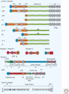
Figure 1.
Figure 1.
Basement membrane proteins and domains. The large glycosylated proteins of the basement membrane consist of tandem repeats of various protein motifs and likely evolved through gene duplication. The laminin subunits consist of arrays that can contain amino-terminal globules (LN domain), laminin-type epidermal growth factor-like repeats that form rodlike regions (each an LE domain that contains four cystine pairs), L4 domains (globule interrupting two half-LE domains such that the second and fourth cysteines bridge across the base of the globule), LF (unique globule of β-subunits), a modified LF domain (here designated LFx), a long coiled-coil domain (a knoblike Lβ subdomain interrupts the heptad-repeats of the β subunits), and terminal LG domains (carboxy-terminal laminin globular domains, each a β-sandwich found in α-subunits). Each laminin consists of an α, β, and γ subunit joined in parallel at the coiled-coil domain and stabilized by disulfide pairs between each subunit at the LE/coiled-coil junction and between the β and γ subunits near the carboxyl terminus. The domain nomenclature shown is as recently revised (Aumailley et al. 2005). Nidogens are laminin-binding proteins that possess their own globular domains (G1-G3) separated by EGF-like domains (generally six cysteines each), thyroglobulin type I repeats (Ty), and a G1-adjacent unique rodlike segment. Netrins possess a laminin-type LN domain, LE repeats, and a unique carboxy-terminal netrin domain. Perlecan consists of five regions consisting of an amino-terminal domain from which project heparan sulfates (HS), SEA domains (sea urchin enterokinase and agrin domain), LDL-receptor repeats, laminin short-arm region consisting of duplicated L4 and LE domains, immunoglobulin (Ig) repeats, and laminin-type LG domains separated by EGF-type repeats. Agrins consist of an amino-terminal laminin binding-domain (NtA), follistatin-like repeats (FS), LE domains, serine/threonine-rich (S/T) domains, and LG domains flanked by EGF-like repeats. Splice variations at two sites impart heparin-binding (A/y insert) and acetylcholine-clustering activity (B/z insert) found in neural agrin. A domain map of the most common type IV collagen heterotrimer (α12α2[IV]) is shown. The triple helical domain, containing multiple interruptions of the gly-x-y repeat (vertical bars), imparts flexibility and is thought to facilitate branching during assembly.
Figure 2.
Figure 2.
Basement membrane components, receptors, and intermolecular binding. Basement membranes contain laminins, nidogens (Nd), type IV collagens, perlecan (perl), and agrins (Ag). Receptors and other cell surface binding molecules include integrins, dystroglycan (DG), the Lutheran glycoprotein (Lu), and sulfated glycolipids such as sulfatides (SGL). Laminin-3A32 is involved in hemidesmosome (HD) assembly, interacting with the α6β4 integrin and BP180. Laminins differ based on their complement of domains, ability to polymerize, proteolytic processing, receptor-binding repertoire, and receptor affinities. Relative strong (heavy solid and dashed lines) and weak (thin dashed lines) interactions are indicated with heavy and thin lines with approximate dissociation constants are indicated where known (small numbers in nM values) (Denzer et al. 1998; Gesemann et al. 1998; Hopf et al. 1999; Talts et al. 1999; Talts et al. 2000; Hopf et al. 2001; Nielsen and Yamada 2001; Ries et al. 2001; Garbe et al. 2002; Smirnov et al. 2002; Nishiuchi et al. 2006; Harrison et al. 2007).
Figure 3.
Figure 3.
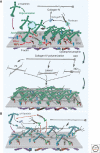
Basement membrane assembly. (A) Steps in the assembly of a basement membrane initiated by a polymerizing laminin. The laminin LG domains bind to a competent cell surface through sulfated glycolipids (SGL), available integrins and α-dystroglycan, promoting laminin polymerization through its LN domains. The α-LN domain also binds to sulfatides and integrins, forcing the laminin onto its side and enabling activation of a new subset of integrins. Nidogens bind to the coiled-coil domain of laminin and to type IV collagen, forming a stabilizing bridge (the collagen also binds to the developing basement membrane through other poorly characterized interactions). Type IV collagen polymerizes to form a second covalently stabilized network. Agrin and perlecan bind to the laminin coiled-coil and to nidogen respectively and also bind to dystroglycan (DG), integrins, and sulfated glycolipids, establishing collateral linkages to additional receptors. Heparin-binding growth factors (GF) bind to the heparan sulfate chains and to their receptor tyrosine kinases (RTK), activating signaling pathways in concert with integrin activation. (B) Steps in the assembly of a basement membrane initiated by a nonpolymerizing laminin. In the case of α4-laminin with weak LG interactions and no α-LN domain, anchorage may depend heavily on collateral linkage through agrin and perlecan. The laminin establishes links to type IV collagen through nidogen; however, in the absence of polymerization, the resulting scaffold has a lower laminin density.

References

    1. Abrahamson DR 1985. Origin of the glomerular basement membrane visualized after in vivo labeling of laminin in newborn rat kidneys. J Cell Biol 100: 1988–2000 - PMC - PubMed
    1. Abrahamson DR 1987. Structure and development of the glomerular capillary wall and basement membrane. Am J Physiol 253: F783–F794 - PubMed
    1. Abrahamson DR, Hudson BG, Stroganova L, Borza DB, St. John PL 2009. Cellular origins of type IV collagen networks in developing glomeruli. J Am Soc Nephrol 20: 1471–1479 - PMC - PubMed
    1. Alcaraz J, Xu R, Mori H, Nelson CM, Mroue R, Spencer VA, Brownfield D, Radisky DC, Bustamante C, Bissell MJ 2008. Laminin and biomimetic extracellular elasticity enhance functional differentiation in mammary epithelia. EMBO J 27: 2829–2838 - PMC - PubMed
    1. Aumailley M, Bruckner-Tuderman L, Carter WG, Deutzmann R, Edgar D, Ekblom P, Engel J, Engvall E, Hohenester E, Jones JC, et al. 2005. A simplified laminin nomenclature. Matrix Biol 24: 326–332 - PubMed

Publication types

LinkOut - more resources