Review
. 2011 Mar 1;3(3):a004838.
doi: 10.1101/cshperspect.a004838.

Not just fat: the structure and function of the lipid droplet

Affiliations
Review

Not just fat: the structure and function of the lipid droplet

Toyoshi Fujimoto et al. Cold Spring Harb Perspect Biol. .

Abstract

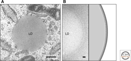
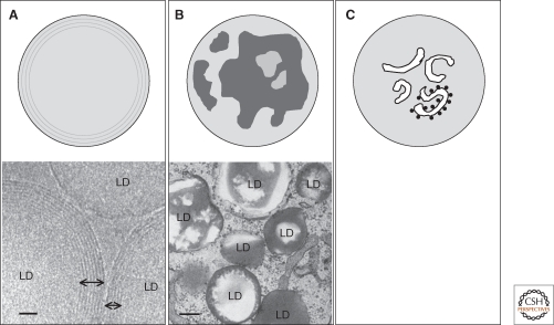
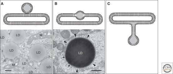
Lipid droplets (LDs) are independent organelles that are composed of a lipid ester core and a surface phospholipid monolayer. Recent studies have revealed many new proteins, functions, and phenomena associated with LDs. In addition, a number of diseases related to LDs are beginning to be understood at the molecular level. It is now clear that LDs are not an inert store of excess lipids but are dynamically engaged in various cellular functions, some of which are not directly related to lipid metabolism. Compared to conventional membrane organelles, there are still many uncertainties concerning the molecular architecture of LDs and how each function is placed in a structural context. Recent findings and remaining questions are discussed.


Figures

Figure 1.
Figure 1.
The current paradigm of the LD structure. (A) Conventional electron microscopy. LDs are observed as spherical structures with no internal structure and are not delimited by the conventional unit membrane. Bar, 200 nm. (B) Cryoelectron microscopy of the isolated LD. The LD surface is seen as a single electron-dense line, which is probably derived from a row of phosphorus atoms in the phospholipid monolayer. Bar, 20 nm.
Figure 2.
Figure 2.
Possible structures in the LD core. (A) Concentric lipid ester layers. Cholesterol ester was proposed to make concentric layers in the LD core. The concentric lines observed by cryoelectron microscopy (shown by bidirectional arrows) may correspond to the cholesterol ester layer boundaries. Bar, 20 nm. (B) Irregular partitioning of lipid esters. The electron micrograph shows that cholesterol esters (low electron density) and triglycerides (high electron density) are segregated in LDs. In this example, cells were sequentially loaded with cholesterol and docosahexanoic acid (Cheng et al. 2009). Bar, 200 nm. (C) Internal membrane-like structures in the core. The membranous structures are occasionally studded with ribosomes suggesting their ER origin.
Figure 3.
Figure 3.
Possible relationship of LDs and the ER. (A) Close apposition. LDs and the ER (*) are often located closely and may make membrane contacts. Mitochondria (M) are also frequently adjacent to LDs. Bar, 200 nm. (B) Intercalation. Huh7 cells loaded with docosahexanoic acid show large LDs intercalated in the ER membrane (arrowheads). The arrows indicate the points in which the ER membrane is continuous with an LD. Bar, 200 nm. (C) Connection by a membrane stalk. The stalk may be a conduit of lipid esters between the LD and the ER membrane.

References

    1. Aboulaich N, Ortegren U, Vener AV, Stralfors P 2006. Association and insulin regulated translocation of hormone-sensitive lipase with ptrf. Biochem Biophys Res Commun 350: 657–661 - PubMed
    1. Agarwal AK, Arioglu E, De Almeida S, Akkoc N, Taylor SI, Bowcock AM, Barnes RI, Garg A 2002. Agpat2 is mutated in congenital generalized lipodystrophy linked to chromosome 9q34. Nat Genet 31: 21–23 - PubMed
    1. Austin JR II, Frost E, Vidi PA, Kessler F, Staehelin LA 2006. Plastoglobules are lipoprotein subcompartments of the chloroplast that are permanently coupled to thylakoid membranes and contain biosynthetic enzymes. Plant Cell 18: 1693–1703 - PMC - PubMed
    1. Baba M, Osumi M, Ohsumi Y 1995. Analysis of the membrane structures involved in autophagy in yeast by freeze-replica method. Cell Struct Funct 20: 465–471 - PubMed
    1. Barba I, Chavarria L, Ruiz-Meana M, Mirabet M, Agullo E, Garcia-Dorado D 2009. Effect of intracellular lipid droplets on cytosolic CA2+ and cell death during ischaemia-reperfusion injury in cardiomyocytes. J Physiol 587: 1331–1341 - PMC - PubMed

Publication types