. 2011 Dec;21(12):1554-69.
doi: 10.1093/glycob/cwr028. Epub 2011 Mar 18.

Conserved oligomeric Golgi complex specifically regulates the maintenance of Golgi glycosylation machinery

Affiliations

Conserved oligomeric Golgi complex specifically regulates the maintenance of Golgi glycosylation machinery

Irina D Pokrovskaya et al. Glycobiology. 2011 Dec.

Abstract

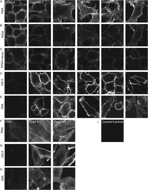
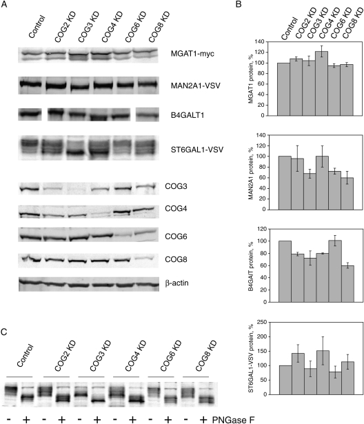
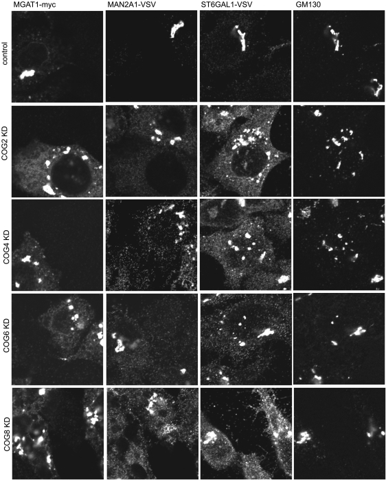
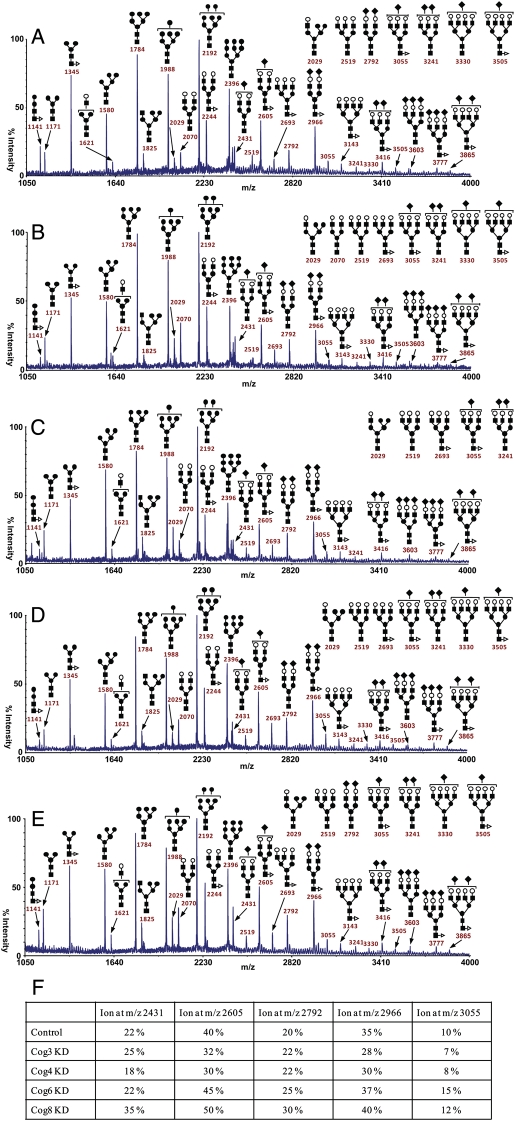
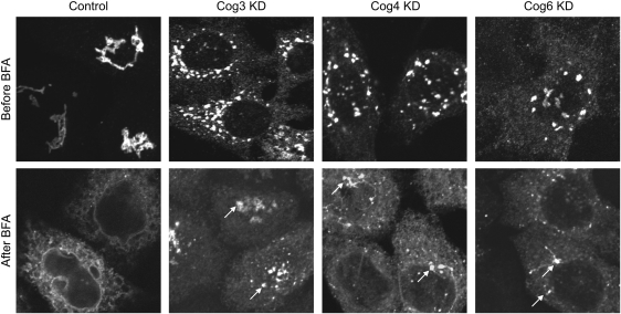
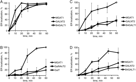
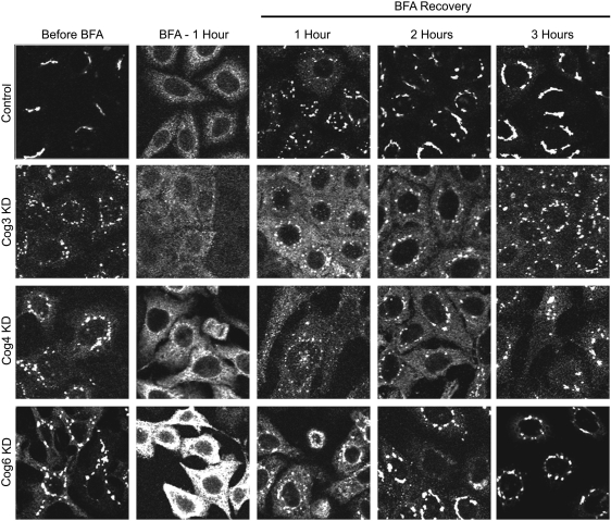
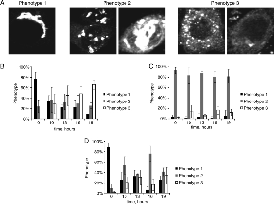
Cell surface lectin staining, examination of Golgi glycosyltransferases stability and localization, and matrix-assisted laser desorption ionization-time of flight (MALDI-TOF) analysis were employed to investigate conserved oligomeric Golgi (COG)-dependent glycosylation defects in HeLa cells. Both Griffonia simplicifolia lectin-II and Galanthus nivalus lectins were specifically bound to the plasma membrane glycoconjugates of COG-depleted cells, indicating defects in activity of medial- and trans-Golgi-localized enzymes. In response to siRNA-induced depletion of COG complex subunits, several key components of Golgi glycosylation machinery, including MAN2A1, MGAT1, B4GALT1 and ST6GAL1, were severely mislocalized. MALDI-TOF analysis of total N-linked glycoconjugates indicated a decrease in the relative amount of sialylated glycans in both COG3 KD and COG4 KD cells. In agreement to a proposed role of the COG complex in retrograde membrane trafficking, all types of COG-depleted HeLa cells were deficient in the Brefeldin A- and Sar1 DN-induced redistribution of Golgi resident glycosyltransferases to the endoplasmic reticulum. The retrograde trafficking of medial- and trans-Golgi-localized glycosylation enzymes was affected to a larger extent, strongly indicating that the COG complex regulates the intra-Golgi protein movement. COG complex-deficient cells were not defective in Golgi re-assembly after the Brefeldin A washout, confirming specificity in the retrograde trafficking block. The lobe B COG subcomplex subunits COG6 and COG8 were localized on trafficking intermediates that carry Golgi glycosyltransferases, indicating that the COG complex is directly involved in trafficking and maintenance of Golgi glycosylation machinery.


Figures

Fig. 1.
Fig. 1.
Lectin staining of COG complex-depleted HeLa cells reveals altered glycosylations of plasma membrane glycoconjugates. HeLa cells were mock-transfected or transfected with siRNA to COG2, COG3, COG4, COG6 and COG8. Seventy two hours after transfection, the cells were chilled to 4°C, stained with PNA-rhodamine, WGA-fluorescein, GS-II-Alexa 594 or GNL-Alexa 647 for 30 min at 4°C, fixed with 4% paraformaldehyde, and visualized by fluorescent microscopy (AB, DE, respectively). (C) Cells were treated with neuraminidase before WGA-fluorescein binding. CHO, ldlB (COG1 KO) and ldlC (COG2 KO) cells were chilled to 4°C, stained with PNA-rhodamine, GS-II-Alexa 594 or GNL-Alexa 647 conjugate for 30 min at 4°C, fixed in 4% paraformaldehyde and visualized by fluorescent microscopy (FH, respectively). (I) PNA-rhodamine binding to control HeLa cells was performed in the presence of 10 mM lactose. Samples were viewed by dual-laser confocal microscopy (63× oil objective). Bar, 10 μm.
Fig. 2.
Fig. 2.
Stability of Golgi enzymes in COG KD cells. HeLa cells that stably express VSV or myc-tagged Golgi enzymes were mock-transfected or transfected with siRNA to COG2, COG3, COG4, COG6 and COG8. Seventy two hours after transfection, cells were collected and lysed in hot 2% SDS. Proteins from cell lysates were separated on SDS–PAGE and western blotted with antibody as indicated (A). Experiment was repeated at least three times and protein expression data were quantified. Actin expression was used as a standard (B). (C) Lysates of HeLa cells that stably express ST6GAL1-VSV were incubated with buffer or treated with PNGase F (New England Biolab) overnight before the WB with anti-VSV antibodies.
Fig. 3.
Fig. 3.
Localization of Golgi enzymes in COG KD cells. HeLa cells that stably express VSV or myc-tagged Golgi enzymes were mock-transfected or transfected with siRNA to COG2, COG4, COG6 and COG8. Seventy two hours after transfection, cells were fixed, stained with antibodies as indicated and analyzed by laser confocal fluorescent microscopy.
Fig. 4.
Fig. 4.
MALDI-TOF analysis of N-linked glycoconjugates isolated from COG-deficient HeLa cells. Mass spectrometry results of total cellular N-linked glycoconjugates of control (A), COG3 KD (B), COG4 KD (C), COG6 KD (D) and COG8 KD (E). Analysis was repeated at least two times for each knockdown. (F) Quantification of these results shown in panels A–E. The structural assignments were made based on the MALDI mass and MS-MS experiments. A minor portion of the monofucosylated glycans carries fucose on an antenna rather than the core. Symbols: galactose, open circles; mannose, closed circles; GlcNAc, closed squares; fucose, open triangles; NeuAc, closed diamonds.
Fig. 5.
Fig. 5.
COG complex-depleted cells are defective in the BFA-induced Golgi redistribution to the ER. GALNT2-GFP HeLa cells were mock-transfected or transfected with siRNA to COG3, COG4 and COG6. Seventy two hours after transfection, cells were fixed (before BFA panel) or were exposed to 0.25 µg/mL BFA for 1 h, fixed and analyzed by fluorescent microscopy (after BFA panel). White arrows show remnants of the Golgi apparatus for the COG subunit knockdown cells (63× oil objective). Bar, 10 μm.
Fig. 6.
Fig. 6.
BFA-induced relocalization of trans-Golgi enzymes is specifically blocked in COG complex-deficient cells. GALNT2-GFP and MGAT1-myc HeLa cells were mock-transfected or transfected with siRNA to COG3, COG4 and COG6. Seventy two hours after transfection, cells were exposed to 0.25 µg/mL BFA for 0-, 10-, 20-, 30-, 45- and 60-min, fixed, stained for MGAT1-myc and GalT, and analyzed by fluorescent microscopy. Images were quantified by counting cells that show a complete redistribution of Golgi enzymes to the ER. Four images were captured with the 63× oil objective on a laser confocal microscope for each time point. The mean result of three independent experiments using control cells (A), COG3 KD cells (B), COG4 KD cells (C) and COG6 KD cells (D) was quantified. Error bars represent standard deviations between three independent experiments.
Fig. 7.
Fig. 7.
COG complex knockdown cells are not deficient in Golgi reassembly assay. MGAT1-myc HeLa cells were mock-transfected or transfected with siRNA to COG3, COG4 and COG6. Seventy two hours after transfection, cells were exposed to BFA (0.25 µg/mL) for 1 h and then allowed to recover in fresh 10% FBS medium for times indicated. The cells were fixed before BFA application, 60 min after BFA and 0-, 1-, 2- and 3-h for recovery, permeabilized with Triton X-100, stained with anti-myc antibodies and analyzed by IF microscopy (63× oil objective). Bar, 10 μm.
Fig. 8.
Fig. 8.
Sar1p T39N-induced disassembly of the Golgi is delayed in COG complex-deficient cells. GALNT2-GFP HeLa cells were mock-transfected or transfected with siRNA to COG3 and COG6. Seventy two hours after transfection, the cells were transfected with plasmid expressing Sar1p T39N. Transfected cells were fixed 19 h later and analyzed by IF microscopy using 63× oil objective. Three phenotypes of localization of GALNT2-GFP were observed: phenotype 1 has a normal juxtanuclear or dilated Golgi localization. Phenotype 2 has a punctate distribution of the enzyme present in both the Golgi and ER localization. Phenotype 3 has small vesicles or ER localization of the enzyme (A). Bar, 10 μm. Graphs showing redistribution of GALNT2-GFP from the Golgi apparatus to the ER in control cells (B), COG3 KD cells (C) and COG6 KD cells (D). For statistical analysis, three individuals blindly classified the phenotypes. Error bars represent standard deviations between each individual's phenotype classifications (n > 50).
Fig. 9.
Fig. 9.
Vesicles that carry MGAT1 contain lobe B COG complex subunits. RFP-COG6 and COG6-GFP plasmids (A, B), RFP-COG6 and COG8-GFP (C, D) or RFP-COG6 (E, F) were cotransfected in MGAT1-myc-myc HeLa cells. After 48 h, cells were fixed, stained with anti-myc and/or anti-COG3 antibodies and developed using HyLite-647-conjugated anti-rabbit antibodies, HyLite-647-conjugated anti-mouse antibodies and/or HyLite-488-conjugated anti-mouse antibodies. Cells were viewed by dual-laser confocal microscopy (63× oil objective). RFP-COG6, COG6-GFP and COG8-GFP positively colocalize with the MGAT1-myc on large Golgi cisternae as well as small vesicle-like membranes (red arrows). Staining with endogenous COG3 reveal no colocalization with the RFP-COG6/MGAT1-myc-positive vesicle structures.

References

    1. Aoki D, Lee N, Yamaguchi N, Dubois C, Fukuda MN. Golgi retention of a trans-Golgi membrane protein, galactosyltransferase, requires cysteine and histidine residues within the membrane-anchoring domain. Proc Natl Acad Sci USA. 1992;89:4319–4323. doi:10.1073/pnas.89.10.4319. - DOI - PMC - PubMed
    1. Bonifacino JS, Glick BS. The mechanisms of vesicle budding and fusion. Cell. 2004;116:153–166. doi:10.1016/S0092-8674(03)01079-1. - DOI - PubMed
    1. Bruinsma P, Spelbrink RG, Nothwehr SF. Retrograde transport of the mannosyltransferase Och1p to the early Golgi requires a component of the COG transport complex. J Biol Chem. 2004;279:39814–39823. doi:10.1074/jbc.M405500200. - DOI - PubMed
    1. Chen R, Jiang X, Sun D, Han G, Wang F, Ye M, Wang L, Zou H. Glycoproteomics analysis of human liver tissue by combination of multiple enzyme digestion and hydrazide chemistry. J Proteome Res. 2009;8:651–661. doi:10.1021/pr8008012. - DOI - PubMed
    1. Foulquier F. COG defects, birth and rise! Biochim Biophys Acta. 2009;1792:896–902. - PubMed

Publication types

Substances