. 2011 Jun;79(6):2470-80.
doi: 10.1128/IAI.00375-10. Epub 2011 Mar 21.

MyD88 signaling is required for efficient innate and adaptive immune responses to Paracoccidioides brasiliensis infection

Affiliations

MyD88 signaling is required for efficient innate and adaptive immune responses to Paracoccidioides brasiliensis infection

Flávio V Loures et al. Infect Immun. 2011 Jun.

Abstract

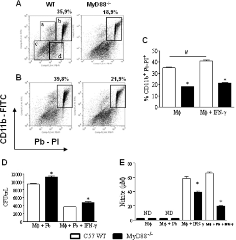
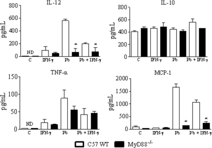
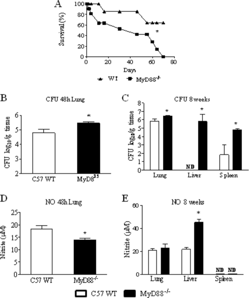
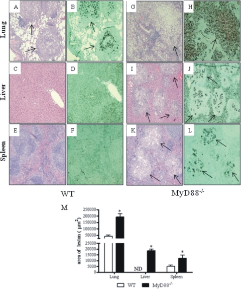
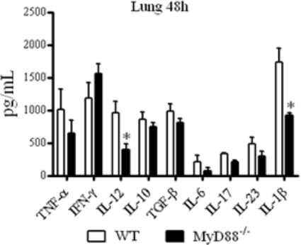
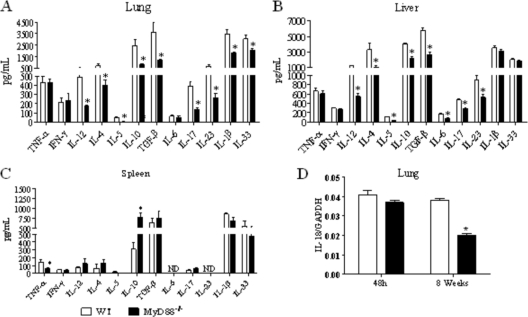
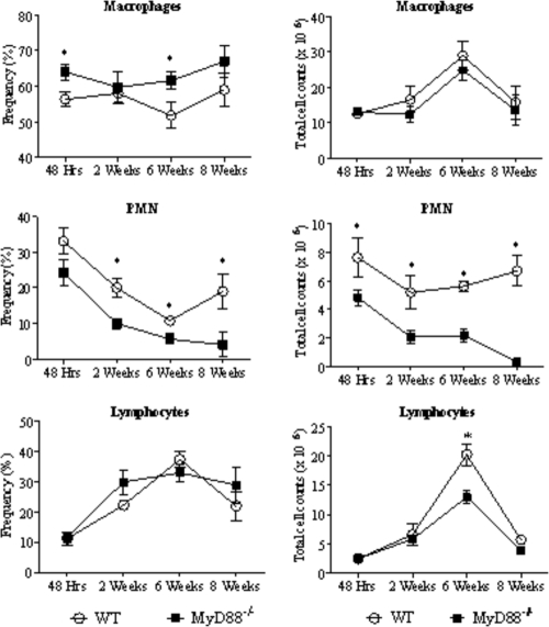
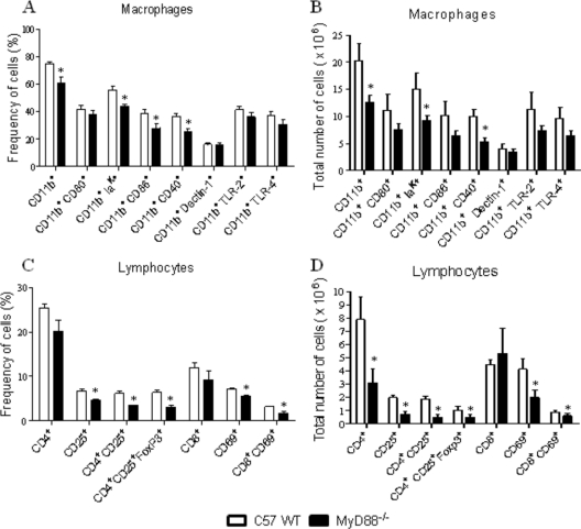
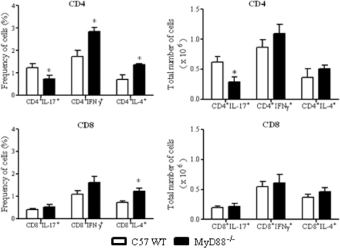
The mechanisms that govern the initial interaction between Paracoccidioides brasiliensis, a primary dimorphic fungal pathogen, and cells of the innate immunity need to be clarified. Our previous studies showed that Toll-like receptor 2 (TLR2) and TLR4 regulate the initial interaction of fungal cells with macrophages and the pattern of adaptive immunity that further develops. The aim of the present investigation was to assess the role of MyD88, an adaptor molecule used by TLRs to activate genes of the inflammatory response in pulmonary paracoccidioidomycosis. Studies were performed with normal and MyD88(-/-) C57BL/6 mice intratracheally infected with P. brasiliensis yeast cells. MyD88(-/-) macrophages displayed impaired interaction with fungal yeast cells and produced low levels of IL-12, MCP-1, and nitric oxide, thus allowing increased fungal growth. Compared with wild-type (WT) mice, MyD88(-/-) mice developed a more severe infection of the lungs and had marked dissemination of fungal cells to the liver and spleen. MyD88(-/-) mice presented low levels of Th1, Th2, and Th17 cytokines, suppressed lymphoproliferation, and impaired influx of inflammatory cells to the lungs, and this group of cells comprised lower numbers of neutrophils, activated macrophages, and T cells. Nonorganized, coalescent granulomas, which contained high numbers of fungal cells, characterized the severe lesions of MyD88(-/-) mice; the lesions replaced extensive areas of several organs. Therefore, MyD88(-/-) mice were unable to control fungal growth and showed a significantly decreased survival time. In conclusion, our findings demonstrate that MyD88 signaling is important in the activation of fungicidal mechanisms and the induction of protective innate and adaptive immune responses against P. brasiliensis.


Figures

Fig. 1.
Fig. 1.
Macrophages from MyD88−/− mice have a decreased ability to interact with P. brasiliensis yeast cells. For phagocytic assays, unprimed (A, C) and IFN-γ-primed (20 ng/ml, overnight) (B, C) peritoneal macrophages from MyD88−/− and WT C57BL/6 mice were infected with 1 × 10⁶/well heat-killed PI-labeled yeast cells (1:1 fungus/macrophage ratio). After 2 h, the supernatants were aspirated, the cells washed, and harvested macrophages labeled with FITC anti-CD11b antibodies and analyzed by flow cytometry. Four different cell populations were identified (regions a to d). (a) Single positive FITC-labeled cells (CD11b+ cells that do not phagocytose or adhere to Pb-PI); (b) double-positive cells (PI-Pb-FITC macrophages); (c) dead cells; and (d) nonphagocytosed PI-Pb. For fungicidal assays, IFN-γ-primed and unprimed peritoneal macrophages were infected with P. brasiliensis yeast cells at a macrophage/yeast ratio of 25:1. After 48 h of cocultivation, the supernatants were obtained to characterize NO and cytokine production; monolayers were lysed and assayed for the presence of viable yeast cells by a CFU assay (D). Supernatants were used to determine the levels of nitrites using the Griess reagent (E). Data are means ± standard errors of the means (SEM) of results from quintuplicate samples from one experiment representative of three independent determinations. # and *, P < 0.05.
Fig. 2.
Fig. 2.
Macrophages from MyD88−/− mice secrete diminished levels of IL-12 and MCP-1. IFN-γ-treated (20 ng/ml) or untreated macrophages of MyD88−/− and WT mice were challenged with viable P. brasiliensis yeast cells (1:25 fungus/macrophage ratio) and cultivated for 48 h at 37°C in 5% CO2. Supernatants were then obtained and used for cytokine measurements using ELISA. Data are means ± SEM of results from triplicate samples from one experiment representative of 3 independent determinations. *, P < 0.05.
Fig. 3.
Fig. 3.
Absence of MyD88 signaling increases mortality rates and tissue fungal burdens. (A) Survival times of MyD88−/− (n = 9) and WT (n = 10) mice after i.t. infection with 1 × 10⁶ P. brasiliensis yeast cells was determined in a period of 70 days. The results are representative of two independent experiments. *, P < 0.05. (B and C) Recovery of fungal loads (CFU) from organs of MyD88−/− and WT mice. The bars represent mean ± SEM log10 numbers of CFU obtained from groups of 6 to 8 mice at 48 h (B) and 8 weeks (C) after infection. Levels of NO (μM) present in tissue homogenates obtained 48 h (D) and 8 weeks (E) after fungal infection. The results are representative of three experiments with equivalent results. *, P < 0.05.
Fig. 4.
Fig. 4.
Photomicrographs of lesions of WT (A to F) and MyD88−/− (G to L) mice at week 8 of infection with 1 × 10⁶ P. brasiliensis yeast cells. Compared with those of MyD88−/− mice (G), the pulmonary lesions of WT mice (A) were smaller (arrows) and composed of organized granulomas containing lower numbers of yeast cells (B). The pulmonary lesions of MyD88−/− mice were composed of confluent, necrotic, unorganized granulomas of various sizes (G) containing an elevated number of fungal cells (arrow in H) and replaced almost all the normal tissue (G, H). The livers (C, D) and spleens (E, F) of WT mice presented a normal morphology; in contrast, the livers (I, J) and spleens (K, L) of MyD88−/− mice presented extensive necrotic lesions (arrows in I and K) containing an elevated number of yeast cells (arrows in panels J and L) surrounded by mononuclear inflammatory exudates. H&E (A, C, E, G, I, K)- and Groccot (B, D, F, H, J, L)-stained lesions (magnification, ×100). *, P < 0.05. (M) Total area of lesions in the lungs, livers and spleen of mice (n = 6) at week 8 after infection. *, P < 0.05.
Fig. 5.
Fig. 5.
Early after infection, lung homogenates of MyD88−/− mice presented decreased levels of IL-12 and IL-1β. MyD88−/− and WT mice were infected with 1 × 10⁶ yeast cells of P. brasiliensis, and 48 h later, lungs were collected and disrupted in 5.0 ml of PBS, and supernatants were analyzed for cytokine content by capture ELISA. The bars depict means ± SEM of cytokine levels (6 to 8 animals per group). The results are representative of two independent experiments. *, P < 0.05.
Fig. 6.
Fig. 6.
At week 8 after infection, organs from MyD88−/− mice presented decreased levels of Th1, Th2, and Th17 cytokines. Lung (A), liver (B), and spleen (C) homogenates of MyD88−/− mice presented decreased levels of cytokines. At week 8 after i.t. infection with 1 × 10⁶ yeast cells of P. brasiliensis, organs from MyD88−/− and WT mice were collected and disrupted in 5.0 ml of PBS and supernatants analyzed for cytokine content by capture ELISA. The bars depict means ± SEM of cytokine levels (6 to 8 animals per group). The results are representative of three independent experiments. *, P < 0.05. (D) Quantitative PCR analysis of IL-18 expression in the lungs of P. brasiliensis-infected WT and MyD88−/− mice. Total lung RNA was obtained, reverse transcribed, and cDNA amplified. Real-time PCR was performed using TaqMan universal master mix. Amplified products were normalized to the amount of GAPDH products. Data represent the means ± SEM of results from two independent experiments.
Fig. 7.
Fig. 7.
MyD88 deficiency determines a decreased recruitment of PMN cells to the lungs. Frequency (left side panels) and absolute number (right panels) of leukocyte subsets (macrophages, PMN neutrophils, and lymphocytes) in the lung-infiltrating leukocytes (LIL) from MyD88−/− and WT mice inoculated i.t. with 1 million P. brasiliensis yeast cells. At different postinfection periods (48 h, 2, 6, and 8 weeks), lungs of both mouse strains (n = 6 to 8) were excised, washed in PBS, minced, and digested enzymatically. Lung cell suspensions were obtained and cytospun onto glass slides. Cells were stained by the Diff-Quik bloodstain. Data are expressed as means ± SEM. *, P < 0.05.
Fig. 8.
Fig. 8.
Decreased numbers of activated macrophages, T lymphocytes, and regulatory T cells were detected in the lungs of MyD88−/− mice at week 8 of infection. Characterization of leukocyte subsets by flow cytometry in the lung-infiltrating leukocytes (LIL) from MyD88−/− and WT mice inoculated i.t. with 1 × 10⁶ P. brasiliensis yeast cells. At week 8 after infection, lungs of both mouse strains (n = 6 to 8) were excised and digested enzymatically. Cell suspensions were obtained and stained as described in Materials and Methods. The stained cells were analyzed immediately with FACSCanto equipment gating on macrophages or lymphocytes, as judged from forward and side light scatters. Twenty thousand cells were counted, and the data are expressed as percentage and absolute number of positive cells. For characterization of Treg cells (CD4+ CD25+ FoxP3+), surface staining of CD25+ and intracellular FoxP3 expression were back-gated on the CD4+ T cell population. Data are expressed as means ± SEM and are representative of two independent experiments. *, P < 0.05.
Fig. 9.
Fig. 9.
Decreased numbers of CD4+IL-17+ cells were detected in the lungs of MyD88−/− mice. Groups (n = 6 or 7) of WT and MyD88−/− mice were infected with 1 × 10⁶ P. brasiliensis yeast cells. The presence of IL-17+, IFN-γ+, and IL-4+ CD4+ and CD8+ T cells in the lung-infiltrating leukocytes was assessed by intracellular cytokine staining by flow cytometry at week 8 after infection. Lung cells were restimulated in vitro with phorbol myristate acetate (PMA)-ionomycin for 6 h and subjected to intracellular staining for IL-17, IL-4, and IFN-γ. The lymphocyte population was gated by the forward/side scatters. The results are from one experiment and are representative of two independent experiments. *, P < 0.05.

Similar articles

Cited by

References

    1. Adachi O., et al. 1998. Targeted disruption of the MyD88 gene results in loss of IL-1- and IL-18-mediated function. Immunity 9:143–150 - PubMed
    1. Biondo C., et al. 2005. MyD88 and TLR2, but not TLR4, are required for host defense against Cryptococcus neoformans. Eur. J. Immunol. 35:870–878 - PubMed
    1. Bonfim C. V., Mamoni R. L., Blotta M. H. 2009. TLR-2, TLR-4 and dectin-1 expression in human monocytes and neutrophils stimulated by Paracoccidioides brasiliensis. Med. Mycol. 47:722–733 - PubMed
    1. Bonifazi P., et al. 2009. Balancing inflammation and tolerance in vivo through dendritic cells by the commensal Candida albicans. Mucosal Immunol. 2:362–374 - PubMed
    1. Bretz C., et al. 2008. MyD88 signaling contributes to early pulmonary responses to Aspergillus fumigatus. Infect. Immun. 76:952–958 - PMC - PubMed

Publication types

MeSH terms

LinkOut - more resources