. 2011 Jun;55(6):2714-28.
doi: 10.1128/AAC.01383-10. Epub 2011 Mar 21.

Decrease in penicillin susceptibility due to heat shock protein ClpL in Streptococcus pneumoniae

Affiliations

Decrease in penicillin susceptibility due to heat shock protein ClpL in Streptococcus pneumoniae

Thao Dang-Hien Tran et al. Antimicrob Agents Chemother. 2011 Jun.

Abstract

Antibiotic resistance and tolerance are increasing threats to global health as antibiotic-resistant bacteria can cause severe morbidity and mortality and can increase treatment cost 10-fold. Although several genes contributing to antibiotic tolerance among pneumococci have been identified, we report here that ClpL, a major heat shock protein, could modulate cell wall biosynthetic enzymes and lead to decreased penicillin susceptibility. On capsular type 1, 2, and 19 genetic backgrounds, mutants lacking ClpL were more susceptible to penicillin and had thinner cell walls than the parental strains, whereas a ClpL-overexpressing strain showed a higher resistance to penicillin and a thicker cell wall. Although exposure of Streptococcus pneumoniae D39 to penicillin inhibited expression of the major cell wall synthesis gene pbp2x, heat shock induced a ClpL-dependent increase in the mRNA levels and protein synthesized by pbp2x. Inducible ClpL expression correlated with PBP2x expression and penicillin susceptibility. Fractionation and electron micrograph data revealed that ClpL induced by heat shock is localized at the cell wall, and the ΔclpL showed significantly reduced net translocation of PBP2x into the cell wall. Moreover, coimmunoprecipitation with either ClpL or PBP2x antibody followed by reprobing with ClpL or PBP2x antibody showed an interaction between ClpL and PBP2x after heat stress. This interaction was confirmed by His tag pulldown assay with either ClpLHis₆ or PBP2xHis₆. Thus, ClpL stabilized pbp2x expression, interacted with PBP2x, and facilitated translocation of PBP2x, a key protein of cell wall synthesis process, contributing to the decrease of antibiotic susceptibility in S. pneumoniae.


Figures
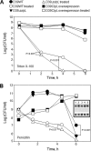
Fig. 1.
Fig. 1.
Increased resistance of pneumococcus to Triton X-100 and penicillin-triggered lysis by ClpL overexpression. Pneumococci were cultured in THY broth until the OD550 was 0.2. Then, Triton X-100 (A) or penicillin (B) was added to the culture at final concentrations of 0.05% and 0.1 μg/ml, respectively, followed by counting of viable cells. Each data point is the mean value from three independent experiments. (Inset) Expression level of ClpL in D39 WT (row 1), D39 ΔclpL (row 2), and the D39 ClpL-overexpressing strain (row 3) after exposure to 0.1 μg/ml penicillin for 0, 1.5, 3, 4.5, and 6 h (lanes 1, 2, 3, 4, and 5, respectively). Statistical differences were analyzed by Student's t test. Standard deviations are smaller than the symbols. The D39 ClpL-overexpressing strain was only marginally affected by Triton X-100 (A) and was not affected by penicillin (B).
Fig. 2.
Fig. 2.
Heat shock-induced ClpL increases penicillin resistance. Pneumococci were cultured in THY broth at 30°C until the OD550 was 0.2 and then heat shocked at 42°C for 30 min. After heat shock, penicillin (Pen) was added to the culture at a final concentration of 0.2 μg/ml, followed by viable cell counting. The figure shows the standard deviation from five independent experiments. Statistical differences were analyzed by Student's t test. In some cases, standard deviations are smaller than the symbol.
Fig. 3.
Fig. 3.
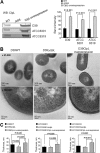
Thicker cell wall by ClpL overexpression. (A) Amount of ClpL in WT, ΔclpL, and ClpL-overexpressing type 2, 1, and 19 strains. WT, ΔclpL, and ClpL-overexpressing strains were grown in THY broth at 37°C until the OD550 was 0.3. Ten micrograms of the cell lysates was applied for Western blot (WB) analysis to determine ClpL level. Colorimetry was used to detect HRP-conjugated secondary antibody used in Western blots. Band density was analyzed by Photoshop. The figure shows representative results of three independent experiments. (B and C) ClpL overexpression increased cell wall thickness. Pneumococci were grown in THY broth until mid-exponential phase (OD550 = 0.3). The bacterial pellet was fixed and used to analyze cell wall thickness by TEM at ×25,000 magnification (upper panels) or ×100,000 magnification (lower panels). TEM images of serotypes 1 and 19 are not shown (B). The figure shows representative results of three independent experiments. Cell wall thickness was determined from 10 random bacterial cells for each strain (C). Significant differences were analyzed by one-way ANOVA.
Fig. 4.
Fig. 4.
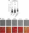
Longer chains and mucoid colonies by ClpL overexpression. Pneumococcal strains at mid-exponential phase (OD550 = 0.3) were fixed onto a glass slide (B, upper panels), and the average chain length was analyzed by microscopy at ×100 magnification from 100 random samples (A and B); or the strains were streaked onto a THY blood agar plate without antibiotic to observe colony morphology (B, lower panels). The figure shows representative results of three independent experiments. Significant differences were analyzed by one-way ANOVA.
Fig. 5.
Fig. 5.
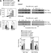
Induction of ClpL by penicillin stress and stabilization of PBP2x expression by ClpL. (A) After exposure of D39 WT to penicillin for 1 h, bacterial RNA was isolated and mRNA levels of clpL, murM, and pbp2x were analyzed by quantitative RT-PCR. Each sample was tested in quadruplicate. The figure shows the standard deviation from three independent experiments. Significant differences were analyzed by one-way ANOVA. (B) D39 WT (upper panels) and D39 ΔclpL (lower panels) at the mid-exponential phase (OD550 = 0.3) were exposed to penicillin for 20 and 60 min. Ten micrograms of bacterial lysate was used for Western blotting (WB) with either anti-ClpL or anti-PBP2x antibody. Both chemiluminescence (for PBP2x) and colorimetry (for ClpL) were used to detect HRP-conjugated secondary antibody used in Western blots. The figure shows results representative of three independent experiments. Band density at 20 min was analyzed by Photoshop. The figure shows standard deviations from three independent experiments.
Fig. 6.
Fig. 6.
Induction of ClpL and PBP2x by heat shock. (A) D39 WT and D39 ΔclpL at the mid-exponential phase (OD550 = 0.3) were heat shocked at 42°C for 30 min. Ten micrograms of bacterial lysate was use to analyze ClpL protein level by Western blot. Colorimetry was used to detect HRP-conjugated secondary antibody used in Western blots. The figure shows representative results of three independent experiments. Band density was analyzed by Photoshop. The figure shows standard deviations from three independent experiments. (B and C) After heat shock, mRNA levels were determined by quantitative RT-PCR (B), and PBP2x protein levels were analyzed by enzyme-linked immunosorbent assay (C) from five (B) and six (C) independent experiments. Significant differences were analyzed by one-way ANOVA.
Fig. 7.
Fig. 7.
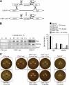
Increase of ClpL expression correlated with PBP2x expression and penicillin susceptibility. (A) An S. pneumoniae D39 clpL-regulated strain (D39 F-clpL) was constructed by placing clpL under the control of the PfcsK inducible promoter in the chromosome of S. pneumoniae. (B) Bacteria were cultured in THY broth until the OD550 was 0.3 and then exposed to 0, 0.1, 0.2, 0.5, and 1% l-fucose for 4 h. Ten micrograms of bacterial lysate was used to analyze the protein expression level of ClpL, PBP2x, and VncS by Western blotting. Band density was analyzed by Photoshop. The figure shows representative results of two independent experiments. (C) D39 WT, D39 ΔclpL, and D39 F-clpL were cultured in THY broth until the OD550 was 0.3. An aliquot of 50 μl of each culture was spread onto a THY agar plate containing 5% sheep blood and 0, 0.1, 0.2, 0.5, or 1% l-fucose. Penicillin (0.02 μg) was placed on sterile filter paper disks, and plates were then incubated at 37°C for 1 day.
Fig. 8.
Fig. 8.
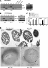
Colocalization of ClpL with PBP2x at the cell wall after heat shock. (A to C) After heat shock, the cell wall (W), cell membrane (M), and cytosol (C) of D39 WT (A) and D39 ΔclpL (B) were fractionated. Western blotting using anti-ClpL, anti-PBP2x, and anti-VncR (cytosole marker) antibodies was carried out to localize ClpL and PBP2x. Purified VncR (VncRHis6), PBP2x (PBP2xHis6), and ClpL (ClpLHis6) were added as positive controls. Total cell lysate was used as input. Both chemiluminescence (for ClpL and VncR) and colorimetry (for PBP2x) were used to detect HRP-conjugated secondary antibody used in Western blots. Band density was analyzed by Photoshop. The figure shows the standard deviations from two independent experiments (C). Significant differences were analyzed by ANOVA. (D) Pneumococci cultured at 30°C were heat shocked at 42°C for 30 min. Thin-sectioned samples were treated with preimmune rabbit serum as primary antibody (left and upper panel) or anti-ClpL antibodies, followed by anti-rabbit IgG conjugated to colloidal gold (right, upper and lower panels). The colloidal gold is seen as electron-dense particles (black arrows). The figure shows representative data from three different sections.
Fig. 9.
Fig. 9.
Interaction of ClpL with PBP2x in vivo and in vitro. (A) Pneumococci cultured at 30°C were heat shocked at 42°C for 30 min. After heat shock, 200 μg of cell lysates of D39 WT was immunoprecipitated (IP) using 0.1 μl (lane 2), 0.5 μl (lane 3), and 1.0 μl (lane 4) of either anti-ClpL (upper panels) or anti-PBP2x (middle panels) antibody. Lysate of the ΔclpL strain was used as a control. The immunoprecipitated products were used for Western blotting (WB) using either anti-ClpL or anti-PBP2x antibody. As a control, preimmune (normal) serum was used instead of anti-ClpL or anti-PBP2x antibody (lower panel), followed by ClpL and PBP2x detection. Purified ClpL and PBP2x were used as positive controls. For the non-heat shock condition (right panels), 1 μl, 2 μl, and 5 μl of either anti-ClpL (upper panels) or anti-PBP2x (lower panels) were used for immunoprecipitation. The figure shows representative results of two independent experiments. (B) Interaction of ClpL and PBP2x in vitro. D39 WT was heat shocked, and the lysate was used for His tag pulldown assay using either ClpLHis6 (left panel) or PBP2xHis6 (right panel). After the membrane was probed with anti-ClpL antibody, the membrane was reprobed with anti-PBP2x antibody, and vice versa. The figure shows representative results of two independent experiments. Chemiluminescence was used to detect HRP-conjugated secondary antibody used in Western blots. GtfAHis6 was used as a negative control.
Fig. 10.
Fig. 10.
Model of how ClpL could increase penicillin resistance. Under heat shock condition, ClpL protein expression is induced, which contributes to the stabilization and increase of PBP2x protein level. As a molecular chaperone, ClpL then interacts with PBP2x in the cytosol and facilitates translocation of PBP2x into the cell wall, which results in a PBP-mediated increase in cell wall thickness and penicillin resistance.

Similar articles

Cited by

References

    1. Andrews J. M. 2001. Determination of minimum inhibitory concentrations. J. Antimicrob. Chemother. 48(Suppl 1):5–16 - PubMed
    1. Avery O. T., MacLeod C. M., McCarty M. 1944. Studies on the chemical nature of the substance inducing transformation of pneumococcal types. Induction of transformation by a desoxyribonucleic acid fraction isolated from pneumococcus type III. J. Exp. Med. 79:137–158 - PMC - PubMed
    1. Balaban N. Q., Merrin J., Chait R., Kowalik L., Leibler S. 2004. Bacterial persistence as a phenotypic switch. Science 305:1622–1625 - PubMed
    1. Barclay M. L., Begg E. J., Chambers S. T. 1992. Adaptive resistance following single doses of gentamicin in a dynamic in vitro model. Antimicrob. Agents Chemother. 36:1951–1957 - PMC - PubMed
    1. Bartlett J. G., et al. 2000. Practice guidelines for the management of community-acquired pneumonia in adults. Clin. Infect. Dis. 31:347–382 - PMC - PubMed

Publication types

MeSH terms

LinkOut - more resources