Review
. 2011 Mar 21;192(6):907-17.
doi: 10.1083/jcb.201009141.

Dynamics of adherens junctions in epithelial establishment, maintenance, and remodeling

Affiliations
Review

Dynamics of adherens junctions in epithelial establishment, maintenance, and remodeling

Buzz Baum et al. J Cell Biol. .

Abstract

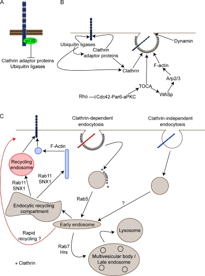
The epithelial cadherin (E-cadherin)-catenin complex binds to cytoskeletal components and regulatory and signaling molecules to form a mature adherens junction (AJ). This dynamic structure physically connects neighboring epithelial cells, couples intercellular adhesive contacts to the cytoskeleton, and helps define each cell's apical-basal axis. Together these activities coordinate the form, polarity, and function of all cells in an epithelium. Several molecules regulate AJ formation and integrity, including Rho family GTPases and Par polarity proteins. However, only recently, with the development of live-cell imaging, has the extent to which E-cadherin is actively turned over at junctions begun to be appreciated. This turnover contributes to junction formation and to the maintenance of epithelial integrity during tissue homeostasis and remodeling.


Figures

Figure 1.
Figure 1.
Factors required to polarize an epithelium. (A) E-Cadherin can dimerize and form trans-homophilic interactions to form cadherin clusters. Ca2+ ions are required to stiffen the extracellular domain and are essential to form homophilic interactions. The E-cadherin intracellular domain contains binding sites for the catenins p120 and β-catenin, thereby forming the cadherin–catenin complex. p120 catenin links cadherin to microtubules and is also important to prevent cadherin endocytosis and degradation. β-Catenin binds α-catenin, which in turn binds actin and several actin-associated proteins, including α-actinin, vinculin, and formin-1. The cadherin–catenin complex also binds many other proteins, including signaling proteins, and cell surface receptors and forms a hub for protein–protein interactions. (B) AJ maturation promotes the assembly of the tight junction (TJ) in vertebrates and the septate junction (SJ) in Drosophila epithelial cells, which both function to provide a paracellular diffusion barrier. The AJ is also necessary to form distinct apical and basolateral domains within the cell with conserved protein complexes that are required to establish and maintain these domains. Apical polarity proteins are highlighted in red; basolateral polarity proteins are highlighted in blue. Apical polarity proteins are found throughout the apical domain but are found concentrated just above the AJ (red boxes). Basolateral proteins are found concentrated just below the AJ (blue boxes), and a mutual inhibition between apical and basolateral complexes maintains this apicobasal polarity. (C) The cytoskeleton is also polarized within epithelial cells and several Rho GTPases, and polarity proteins influence the localization and activity of these cytoskeletal structures (Georgiou and Baum, 2010). Other subcellular structures, although not depicted, are also organized along the apicobasal axis, including the centrosome and the Golgi. Baz, Bazooka. Crb, Crumbs. DaPKC, Drosophila aPKC. DLG, discs large. LGL, lethal giant larvae. Sdt, Stardust. Yrt, yurt.
Figure 2.
Figure 2.
The regulation of E-cadherin recycling. (A) p120 catenin inhibits cadherin endocytosis and degradation by preventing the association of adaptor complexes with the cadherin juxtamembrane intracellular region (Fujita et al., 2002; Ishiyama et al., 2010), which prevents cadherin recruitment into clathrin-coated pits. (B) Dissociation between cadherin and p120 allows adaptors, such as AP-2 and β-arrestin, to recruit clathrin and other accessory proteins to promote internalization. Additionally, specific ubiquitin conjugates (E2) and ligases (E3) may act as adaptors for clathrin or as connectors to AP-2 adaptors to activate the clathrin-coated endocytosis machinery. Cdc42–Par6–aPKC, via TOCA proteins and Arp2/3, promotes dynamin-mediated endocytosis. (C) E-Cadherin can undergo either clathrin-dependent (red) or -independent (blue) endocytosis (Delva and Kowalczyk, 2009), and its possible trafficking routes are depicted here together with several proteins that have been shown to have a demonstrated role in E-cadherin trafficking (Lock and Stow, 2005; Palacios et al., 2005; Bryant et al., 2007; Toyoshima et al., 2007). Both trafficking routes converge onto the Rab5-positive early endosome, which sorts its cargo for recycling or degradation. It is not known whether E-cadherin uses the Rab4-dependent rapid recycling route to facilitate its trafficking.
Figure 3.
Figure 3.
AJ assembly in vitro and in vivo. (A, 1) Cell contact and E-cadherin engagement in vitro leads to a remodeling of the actin cytoskeleton (green), promoting lamellipodial and filopodial protrusions via Rac, Cdc42, and Arp2/3 activity. (2) These dynamic protrusions promote further E-cadherin interactions and clustering. The nascent AJs are connected to the circumferential actomyosin cable via contractile actin bundles (blue). (3) Myosin-mediated contraction expands intercellular contact and aligns cadherin–catenin complexes (red bars), leading to the maturation of the junction. (B) Fusion between epithelial sheets in vivo again shows cooperation between dynamic protrusions (green arrows) and actomyosin cables (blue arrows). (1 and 2) An actomyosin cable assembles at the edge of each epithelial sheet, forcing the two sheets together. (3) Individual cells on the leading edge of each epithelial sheet form filopodia (green) that engage with one another, forming cadherin–catenin clusters at the points of contact (red), which are required to seal the two sheets together. In the case of the Drosophila embryo during dorsal closure, the ectodermal sheets migrate over a squamous epithelium called the amnioserosa. Here, the apical constriction of amnioserosa cells has been shown to promote dorsal closure (inset). Green arrows represent protrusive activity; blue arrows represent contractile activity.
Figure 4.
Figure 4.
Different effects of actomyosin-mediated constriction during tissue remodeling. (A) Cell intercalation requires a polarized redistribution of proteins within the plane of the epithelium to limit remodeling to specific junctions. Before cell intercalation, actin and myosin (blue) as well as E-cadherin, Armadillo/β-catenin, and Bazooka/Par-3 (red) localize uniformly at the cortex. At the onset of cell intercalation, planar symmetry is broken, with F-actin and myosin II concentrating at anteroposterior interfaces (blue). Conversely, Bazooka/Par-3, E-cadherin, and Armadillo/β-catenin accumulate at dorsoventral interfaces (red). This limits actomyosin contractility to the anteroposterior interfaces (blue arrows). (B) The same polarized redistribution of proteins is required to form actomyosin cables that span multiple pairs of cells. Contraction results in the formation of multicellular rosettelike patterns. Blue arrows represent contractile activity. (C) The apical actomyosin medial weblike network (blue) can force apical constriction by shortening all junctions. (D) Basally localized and highly polarized actomyosin parallel bundles (blue) force an oscillating directional constriction at the base of the cell.

References

    1. Abraham S., Yeo M., Montero-Balaguer M., Paterson H., Dejana E., Marshall C.J., Mavria G. 2009. VE-Cadherin-mediated cell-cell interaction suppresses sprouting via signaling to MLC2 phosphorylation. Curr. Biol. 19:668–674 10.1016/j.cub.2009.02.057 - DOI - PubMed
    1. Adams C.L., Chen Y.T., Smith S.J., Nelson W.J. 1998. Mechanisms of epithelial cell–cell adhesion and cell compaction revealed by high-resolution tracking of E-cadherin–green fluorescent protein. J. Cell Biol. 142:1105–1119 10.1083/jcb.142.4.1105 - DOI - PMC - PubMed
    1. Aigouy B., Farhadifar R., Staple D.B., Sagner A., Röper J.C., Jülicher F., Eaton S. 2010. Cell flow reorients the axis of planar polarity in the wing epithelium of Drosophila. Cell. 142:773–786 10.1016/j.cell.2010.07.042 - DOI - PubMed
    1. Akhtar N., Hotchin N.A. 2001. RAC1 regulates adherens junctions through endocytosis of E-cadherin. Mol. Biol. Cell. 12:847–862 - PMC - PubMed
    1. Assémat E., Bazellières E., Pallesi-Pocachard E., Le Bivic A., Massey-Harroche D. 2008. Polarity complex proteins. Biochim. Biophys. Acta. 1778:614–630 10.1016/j.bbamem.2007.08.029 - DOI - PubMed

Publication types