. 2011 Mar 21;192(6):1043-55.
doi: 10.1083/jcb.201010111.

The yeast lipin orthologue Pah1p is important for biogenesis of lipid droplets

Affiliations

The yeast lipin orthologue Pah1p is important for biogenesis of lipid droplets

Oludotun Adeyo et al. J Cell Biol. .

Abstract

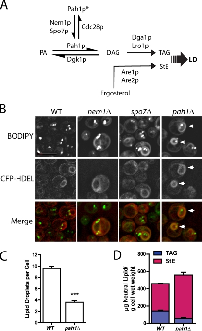
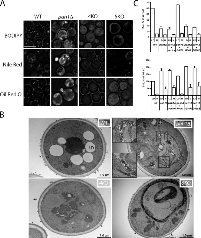
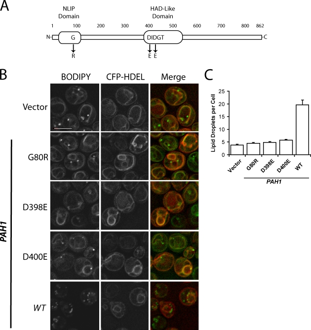
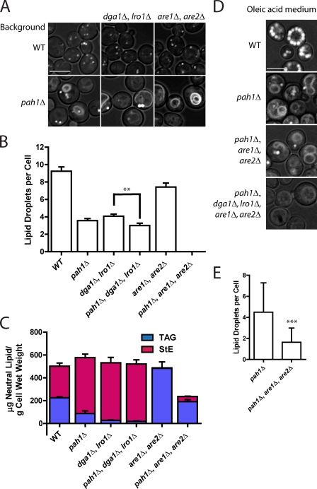
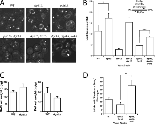
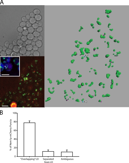
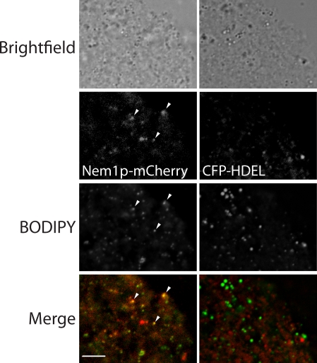
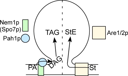
Lipins are phosphatidate phosphatases that generate diacylglycerol (DAG). In this study, we report that yeast lipin, Pah1p, controls the formation of cytosolic lipid droplets. Disruption of PAH1 resulted in a 63% decrease in droplet number, although total neutral lipid levels did not change. This was accompanied by an accumulation of neutral lipids in the endoplasmic reticulum (ER). The droplet biogenesis defect was not a result of alterations in neutral lipid ratios. No droplets were visible in the absence of both PAH1 and steryl acyltransferases when grown in glucose medium, even though the strain produces as much triacylglycerol as wild type. The requirement of PAH1 for normal droplet formation can be bypassed by a knockout of DGK1. Nem1p, the activator of Pah1p, localizes to a single punctum per cell on the ER that is usually next to a droplet, suggesting that it is a site of droplet assembly. Overall, this study provides strong evidence that DAG generated by Pah1p is important for droplet biogenesis.


Figures

Figure 1.
Figure 1.
pah1Δ has fewer lipid droplets but a similar amount of neutral lipid. (A) Abbreviated diagram showing the final steps in neutral lipid synthesis. Pah1p*, phosphorylated cytosolic Pah1p; LD, lipid droplet; TAG, triacylglycerol; StE, steryl ester. (B) BODIPY-stained ER rings (arrows). Fluorescence images of cells of the indicated strains containing the ER marker CFP-HDEL, which were cultured in minimal dextrose medium (SD) and stained with BODIPY. Note the fewer droplets in pah1Δ. Bar, 5 µm. (C) Comparison of droplet number in wild-type (WT) and pah1Δ cells. ***, P < 0.0001; droplets in cells (examining z-section series) from four independent experiments, at least 20 cells per field; SEM shown. (D) Total neutral lipid content from mass spectrometry. Means and ranges are shown from two independent experiments.
Figure 2.
Figure 2.
Neutral lipids accumulate in the ER in the absence of PAH1. (A) BODIPY, Nile red, and Oil red O staining of wild type (WT), pah1Δ, 4KO (dga1Δlro1Δare1Δare2Δ), and 5KO (pah1Δdga1Δlro1Δare1Δare2Δ) strains grown in SD medium. Images incorporate both brightfield and fluorescent channel for the indicated lipophilic dyes. Bar, 5 µm. (B) Inclusions in pah1Δ membranes seen with transmission electron microscopy. The indicated strains were cultured overnight in oleic acid medium before fixation. pah1Δ insets are higher magnifications of boxed areas and are shown to illustrate membrane inclusions. Note the membrane proliferation but no inclusions in the 5KO stain. LD, lipid droplet. (C) Droplet (LD) and membrane (M) levels of TAG and StE in the indicated strains. Postnuclear extracts from spheroplasts (grown in oleate) were fractionated by centrifugation. An identical percentage of the floating droplets and membrane pellets, normalized to total protein in the extracts, was subjected to TLC. Error bars indicate the range of values from two independent experiments.
Figure 3.
Figure 3.
PA phosphatase activity of Pah1p is required for efficient formation of lipid droplets. (A) Diagram of Pah1p primary structure (modified from Han et al., 2007). NLIP, N-terminal lipin homology domain; HAD, haloacid dehalogenase-like domain. (B) The pah1Δ strain expressing the indicated PAH1 alleles on a pRS313 vector. The ER marker CFP-HDEL was expressed on a plasmid and driven by the PGK1 promoter. Lipid droplets were stained with BODIPY. Cells were observed in SD medium. Bar, 5 µm. (C) Droplet number per cell in strains shown in B. Error bars represent SEM based on three fields (∼50 cells per field) from three independent experiments. WT, wild type.
Figure 4.
Figure 4.
Knockout of PAH1 and steryl acyltransferases abolishes lipid droplets in cells grown in glucose medium. (A) BODIPY staining of the indicated strains in SD. DGA1 and LRO1 encode DAG acyltransferases, and ARE1 and ARE2 encode steryl acyltransferases. Images incorporate both brightfield and FITC channel (for lipid droplets). Bar, 5 µm. (B) Lipid droplet number in strains shown in A. Droplets in ∼30 cells were counted in z-section series from three independent experiments (∼90 cells total per strain). Mean ± SEM is shown. Knockout of pah1Δ always resulted in significantly lower droplets, although the significance in only the middle pair is shown for clarity; **, P < 0.001. (C) Quantification of TAG and StE from mass spectrometry analysis for the indicated strains grown in SD. Means and ranges from two independent experiments are shown. The values for wild type (WT) and pah1Δ are also shown in Fig. 1 D. (D) BODIPY staining of the indicated strains grown in medium containing oleic acid. Bar, 5 µm. (E) A comparison of droplet number in pah1Δ and pah1Δare1Δare2Δ grown in oleic acid. Error bars represent standard deviation; ***, P < 0.0001; based on two fields (∼50 cells per field) from three independent experiments.
Figure 5.
Figure 5.
Knockout of DAG kinase leads to more droplets and a bypass in Pah1p function. (A) Knockout of DGK1 increases droplet number independent of TAG. The indicated strains were grown in SD, and lipid droplets were stained with BODIPY. Images incorporate brightfield channel. Arrows illustrate pearls-on-a-string phenotype. Bar, 5 µm. (B) Droplet number increases in dgk1Δ even in the absence of TAG. The inset illustrates the enzymes that catalyze PA-DAG interconversions; the source for the other PA phosphatase activity is unknown. Droplets were counted in at least three random fields, ∼20 cells per field; mean ± SEM is shown. *, P < 0.05; ***, P < 0.0001. (C) Comparison of DAG and PA levels of wild type (WT) and dgk1Δ, showing an increase in DAG and decrease in PA in the mutant. Error bars are SEM based on measurements from three independent experiments. (D) Histogram showing the percentage of cells with pearls-on-a-string lipid droplet phenotype; mean ± SEM (**, P < 0.001) based on three random fields (at least 20 cells per field) from three independent experiments.
Figure 6.
Figure 6.
Nem1p localizes next to lipid droplets on the ER. (A) Nem1p-mCherry was chromosomally expressed at the NEM1 locus. Lipid droplets were stained with BODIPY. (top left) Brightfield image. (bottom left) Projection image of BODIPY-stained cells showing Nem1p-mCherry localization (bottom left inset). The arrowhead is an example of colocalization of CFP-HDEL (ER), BODIPY (lipid droplet), and Nem1p-mCherry. Bars, 5 µm. (right) Three-dimensional reconstruction of the same field of cells. Green, droplets; pink (10 dots), Nem1p within 0.65 µm of a droplet; blue (two dots with arrowheads), Nem1p-mCherry beyond 0.65 µm of a droplet. (B) Juxtaposition of Nem1p-mCherry puncta with lipid droplets (LD). Nem1p-mCherry puncta were counted from seven separate groups of cells and scored for their observed association with lipid droplets. Mean ± SEM was calculated across all seven groups.
Figure 7.
Figure 7.
Colocalization of Nem1p-mCherry and droplets in isolated membranes. Membranes derived from postnuclear supernatant of yeast strains containing chromosomally expressed Nem1p-mCherry (left) or plasmid-expressed 3-phosphoglycerate kinase (PGK) CFP-HDEL (right) were stained with BODIPY. The arrowheads point to three examples of colocalization of Nem1p-mCherry and BODIPY puncta on membranes. Bar, 5 µm.
Figure 8.
Figure 8.
Working model. Droplets are controlled by Pah1p-generated DAG and Are1p/Are2p-generated StEs in normal glucose medium. The dotted line indicates the possibility that these two inputs control two populations of droplets. St, sterol.

References

    1. Agarwal A.K., Garg A. 2006. Genetic basis of lipodystrophies and management of metabolic complications. Annu. Rev. Med. 57:297–311 10.1146/annurev.med.57.022605.114424 - DOI - PubMed
    1. Andersson L., Boström P., Ericson J., Rutberg M., Magnusson B., Marchesan D., Ruiz M., Asp L., Huang P., Frohman M.A., et al. 2006. PLD1 and ERK2 regulate cytosolic lipid droplet formation. J. Cell Sci. 119:2246–2257 10.1242/jcs.02941 - DOI - PubMed
    1. Antonny B., Huber I., Paris S., Chabre M., Cassel D. 1997. Activation of ADP-ribosylation factor 1 GTPase-activating protein by phosphatidylcholine-derived diacylglycerols. J. Biol. Chem. 272:30848–30851 10.1074/jbc.272.49.30848 - DOI - PubMed
    1. Asp L., Kartberg F., Fernandez-Rodriguez J., Smedh M., Elsner M., Laporte F., Bárcena M., Jansen K.A., Valentijn J.A., Koster A.J., et al. 2009. Early stages of Golgi vesicle and tubule formation require diacylglycerol. Mol. Biol. Cell. 20:780–790 10.1091/mbc.E08-03-0256 - DOI - PMC - PubMed
    1. Bacia K., Schwille P., Kurzchalia T. 2005. Sterol structure determines the separation of phases and the curvature of the liquid-ordered phase in model membranes. Proc. Natl. Acad. Sci. USA. 102:3272–3277 10.1073/pnas.0408215102 - DOI - PMC - PubMed

Publication types

MeSH terms