Multispectral diagnostic imaging of the iris in pigment dispersion syndrome
- PMID: 21423031
- PMCID: PMC3153570
- DOI: 10.1097/IJG.0b013e3182120877
Multispectral diagnostic imaging of the iris in pigment dispersion syndrome
Abstract
Purpose: To determine if wavelength selection with near infrared iris imaging may enhance iris transillumination defects (ITDs) in pigment dispersion syndrome.
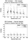
Methods: An experimental apparatus was used to acquire iris images in 6 African-American (AA) and 6 White patients with pigment dispersion syndrome. Light-emitting diode probes of 6 different spectral bands (700 to 950 nm) were used to project light into patients' eyes. Iris patterns were photographed, ITD regions of interest were outlined, and region of interest contrasts were calculated for each spectral band.
Results: Contrasts varied as a function of wavelength (P<0.0001) for both groups, but tended to be highest in the 700 to 800 nm range. Contrasts were higher in Whites than AAs at 700 nm but the opposite was found at 810 nm (P<0.001).
Conclusions: Optimized near infrared iris imaging may be wavelength dependent. Ideal wavelength to image ITDs in more pigmented eyes may be slightly longer than for less pigmented eyes.
Figures


References
-
- Saari M, Vuorre I, Nieminen H. Infrared transillumination stereophotography of normal iris. Can J Ophthalmol. 1977;12:308–311. - PubMed
-
- Alward WLM, Munden PM, Verdick RE, Perell R, Thompson HS. Use of infrared videography to detect and record iris transillumination defects. Arch Ophthalmol. 1990;108:748–750. - PubMed
-
- Haynes WL, Alward WLM, McKinney JK, Munden PM, Verdick R. Quantitation of iris transillumination defects in eyes of patients with pigmentary glaucoma. J Glaucoma. 1994;3:106–113. - PubMed
-
- Roberts DK, Chaglasian MA, Meetz RE. Iris transillumination defects in the pigment dispersion syndrome as detected with infrared videography: a comparison between a group of blacks and a group of nonblacks. Optom & Vis Sci. 1999;76:544–549. - PubMed
Publication types
MeSH terms
Grants and funding
LinkOut - more resources
Full Text Sources
Other Literature Sources
Medical

