. 2011 Apr 20;30(8):1549-62.
doi: 10.1038/emboj.2011.73. Epub 2011 Mar 18.

Ecdysteroids affect Drosophila ovarian stem cell niche formation and early germline differentiation

Affiliations

Ecdysteroids affect Drosophila ovarian stem cell niche formation and early germline differentiation

Annekatrin König et al. EMBO J. .

Abstract

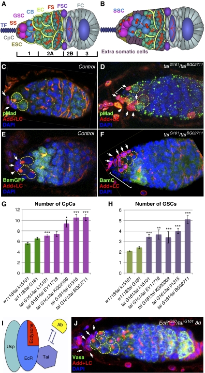
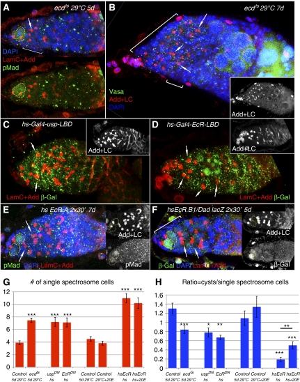
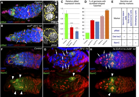
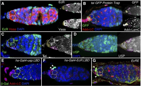
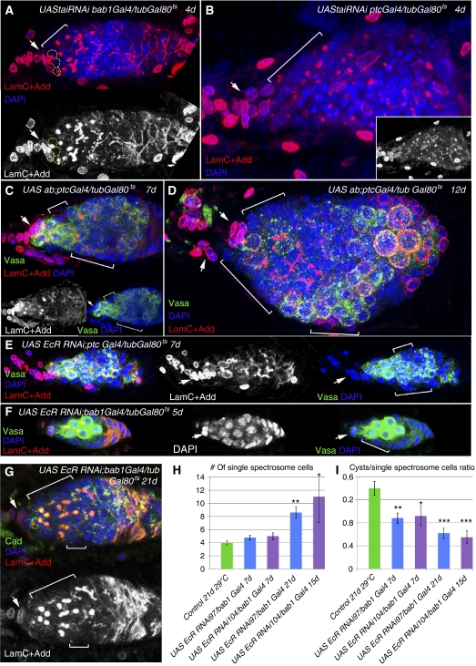
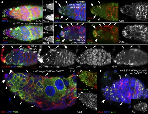
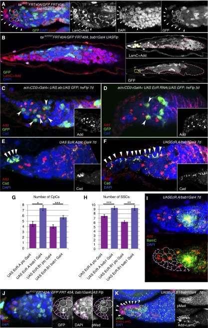
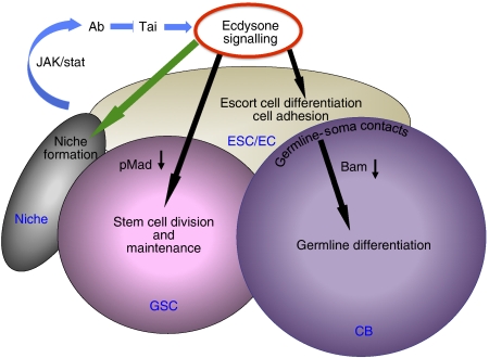
Previously, it has been shown that in Drosophila steroid hormones are required for progression of oogenesis during late stages of egg maturation. Here, we show that ecdysteroids regulate progression through the early steps of germ cell lineage. Upon ecdysone signalling deficit germline stem cell progeny delay to switch on a differentiation programme. This differentiation impediment is associated with reduced TGF-β signalling in the germline and increased levels of cell adhesion complexes and cytoskeletal proteins in somatic escort cells. A co-activator of the ecdysone receptor, Taiman is the spatially restricted regulator of the ecdysone signalling pathway in soma. Additionally, when ecdysone signalling is perturbed during the process of somatic stem cell niche establishment enlarged functional niches able to host additional stem cells are formed.


Conflict of interest statement

The authors declare that they have no conflict of interest.

Figures

Figure 1
Figure 1
The ecdysone receptor co-activator Taiman controls the number of ovarian germline stem cell niche cells. (A) Schematic view of a wild-type germarium: germline stem cells (GSCs, pink) marked by anterior spectrosomes (SS, red dots) are located at the apex of the germarium next to the niche cap cells (CpCs, grey). Further noted are terminal filament (TF; dark blue), escort stem cells (ESCs, olive), differentiating cystoblasts (CBs, blue), escort cells (ECs, lime), 4, 8 (bright green) and 16 cell (green) cysts in region 2A, indicated by the presence of fusomes (FS, red branched structures), follicle stem cells (FSCs, violet) and follicle cells (FC, light grey) in regions 2B and 3. (B) Schematic view of a tai mutant germarium with an increased number of single spectrosome containing cells (SSCs, pink and blue), CpCs (grey) and additional somatic cells (plum). (C, E) In wild-type germaria, two GSCs marked by the presence of the stem cell marker pMad (C), spectrosomes (stained with Adducin) and the absence of the differentiation factor BamC (E) are directly attached to the niche (marked with LaminC, arrows). (D, F) In the tai61G1/taiBG02711 transheterozygous mutant germarium, the enlarged niche is coupled with an increased number of GSCs that are pMad positive (D) and BamC negative (F). In addition, extra somatic cells are present at the anterior (marked with brackets). CpC (G) and GSC (H) numbers are increased in tai mutant germaria. (I) Scheme illustrating that Tai is a co-activator of the EcR/USP nuclear receptor complex that is activated upon binding of its ligand ecdysone; Ab negatively regulates the ecdysone signalling by direct binding to Tai (based on Bai et al (2000) and Jang et al (2009)). (J) EcRQ50st/tai61G1 transheterozygous germaria also contain an increased number of GSCs and CpCs, indicating that tai genetically interacts with EcR (see Supplementary Table S1). (DF, J) Projections of optical sections assembled through the germarial tissue; GSCs are outlined with yellow dashed lines, niche cells are marked with white arrows; Red, Adducin+LaminC; blue, DAPI; and green, pMad (C, D), BamGFP (E), BamC (F) and Vasa (J); Error bars represent s.e.m. *P<0.05, **P<0.005, ***P<0.0005.
Figure 2
Figure 2
Disrupted ecdysone signalling during adulthood results in delayed germline differentiation. (A) At the restrictive temperature (29°C) ecd1ts adult animals contain germaria filled with supernumerary SSCs. (B) Extended depletion of ecdysone furthermore increases the undifferentiated SSC number and causes somatic cell defects affecting cyst pinching off from the germarium. (C, D) Heat shock induced expression of USP and EcR dominant-negative forms (uspDN (hs-Gal-4-usp-LBD) and EcRDN (hs-Gal-4-usp-LBD)) also lead to the appearance of supernumerary SSCs. (E, F) Similarly to the effects that are caused by disturbing the ecdysone pathway via ecd1ts or dominant-negative EcRDN and uspDN mutations, expression of the EcR isoforms EcR.A or EcR.B1 induced by heat shock (twice per day for 30 min 4 days in a row) increases the number of SSCs, but not GSCs and influences CB differentiation. Note the presence of dumbbell-shaped fusomes in (AF). (G) In control conditions around four SSCs per germarium are detected. Ecdysone withdrawal via ecd1ts mutation as well as heat shock-induced expression of uspDN or EcRDN and overexpression of EcR led to a 2- or 2.5-fold increase in SSC number, whereas external supply of ecdysone does not change the amount of SSCs within the germarium. (H) The ratio of differentiating cysts to SSCs is about 1.5-fold decreased in ecd1ts, uspDN and EcRDN mutant germaria. This decrease is even more pronounced (seven times) in hsEcR flies. Providing 20E externally can partially, but significantly alleviate this early germline differentiation delay. (AF) Projections of optical sections assembled through the germarial tissue. GSCs are outlined with yellow dashed lines, dumbbell-shaped fusomes are marked with arrows and additional somatic cells are marked with brackets. Red, LaminC+Adducin; blue, DAPI; and green, pMad (A, E); Vasa (B) and β-galactosidase (C, D, F) Error bars represent s.e.m. *P<0.05, **P<0.005, ***P<0.0005.
Figure 3
Figure 3
Ecdysone signalling affects the TGF-β pathway. (A) Wild-type germarium containing two GSCs, marked by pMad staining. (B) Upon blocked ecdysone production the relative pMad expression levels in GSCs are decreased (C, compare the pMad levels measured by grey value in A and B). (D) Disruption of ecdysone signalling results in the increase of dumbbell-shaped fusome quantity. In ecd1ts4210 mutant flies that were at the restrictive temperature for up to 7 days, 55% (n=33) of the germaria have dumbbell-shaped fusomes (ecd1ts218 51%, n=37) whereas in equally treated w1118 germaria, only 18% (n=11) of the germaria contain dumbbell-shaped fusomes. After overexpression of EcR.A or EcR.B1 for 7 days 67% or 84% (n=15, 19, respectively) of the analysed germaria contain dumbbell-shaped fusomes. (E) The characteristics of GSCs, SSCs and developing cysts are compared schematically. GSCs express the stem cell markers pMad and Dad lacZ and developing cysts the differentiation factor BamC, whereas additional SSCs in germaria deficient of ecdysone signalling are pMad, Dad lacZ and BamC negative, showing that they do not maintain stem cell identity and are delayed in development. (F) In wild-type germarium, BamC is present in developing CBs adjacent to GSCs, while in ecdysone pathway mutants, ecd1ts (G) and hsEcR (H), the anterior part of the germarium is filled with cells that do not express the differentiation marker BamC and contain a single spectrosome or a dumbbell-shaped fusome. (A, B, FH) Projections of optical sections assembled through the germarial tissue. GSCs are outlined with yellow dashed lines, dumbbell-shaped fusomes are marked with arrows and BamC-positive differentiating cysts with arrowheads. Red, Adducin+LaminC; blue, DAPI; and green, pMad (A, B); BamC (FH). Error bars represent s.e.m. Significance calculated using the t-test (C), χ2-test (D). *P<0.05, ***P<0.0005.
Figure 4
Figure 4
Expression pattern of the ecdysone pathway components in the Drosophila germarium. (A) The anti-EcR (common region) antibody detects high levels of EcR in ESCs and FCs. (B) In the tai G00308 protein trap line where GFP is expressed under the control of the endogenous tai promoter, high GFP levels were detected in CpCs, ESCs and FSCs. (C) Comparable expression pattern is observed with the anti-Tai antibody. (D) The nuclear receptor USP detected by the anti-USP antibody shows identical expression pattern to its binding partner EcR. (E, F) Spatial patterns of ecdysone signalling activation identified via β-Gal staining of heat-treated hs-Gal4-usp.LBD/+; UAS-lacZ/+ (E) and hs-Gal4-EcR.LBD/+; UAS-lacZ/+ (D) germaria prove ecdysone signalling being mainly active in the ESCs (marked with arrowheads). (E) The ecdysone signalling reporter EcRE-lacZ shows the presence of active ecdysone transcription complex in ESCs as well (marked with arrowheads). Different cell types are marked as follows: GSCs, white dashed lines; CpCs, yellow dashed lines; ESCs/ECs, red dashed lines; FCs, green dashed lines. Red, Vasa (A), Adducin+LaminC (B, EG); blue, DAPI; and green, anti-EcR (common region) (A); GFP (B); anti-Tai (C), anti-USP (D) and β-galactosidase (EG).
Figure 5
Figure 5
Ecdysteroids act from the soma to regulate the progression of germline development in the germarium. (A, B) The EcR co-activator Tai is downregulated specifically in the somatic cells of the germarium using ptcGal4 and bab1Gal4 in combination with tubGal80ts system to avoid lethality. Upon downregulation of tai in the soma, the number of developmentally delayed SSCs increases dramatically. (C, D) Overexpression of the Tai repressor, Abrupt using UAS ab with the same drivers causes similar phenotypes as seen with downregulation of tai. (B, D) The tai and ab mutant germaria are filled with undifferentiated SSCs, cysts are not pinching off and additional somatic cells (brackets) are in the vicinity. Note the similarity of phenotypes caused by ecdysteriod deficit (ecd1ts, Figure 2B) and disruption of ecdysone signalling pathway components just in germarial soma. (EG) The downregulation of the EcR in the somatic cells of the germarium via expression of UAS EcR RNAi97 under control of ptcGal4 (E) and bab1Gal4 (F, G) leads to an increase of SSCs at the expense of developing cysts. Note the presence of dumbbell-shaped spectrosomes and additional somatic cells. (H) Bar graph showing extra quantities of SSCs upon EcR downregulation via expression of UAS EcR RNAi97 or UAS EcR RNAi104 with the somatic drivers ptcGal4 or bab1Gal4. This phenotype gets more pronounced with longer duration of EcR abolition. (I) The ratio of differentiating cysts to SSCs is also decreased correspondingly to the increase in the SSC number. (AG) Projections of optical sections assembled through the germarial tissue are shown. CpCs are marked with arrows, additional somatic cells with brackets. Red, Adducin+LaminC (AG); blue, DAPI; and green, Vasa (CF), Cadherin (G). Error bars represent s.e.m. *P<0.05, **P<0.005, ***P<0.0005.
Figure 6
Figure 6
Cell adhesion and cytoskeleton proteins are misregulated in escort cells mutant for ecdysone signalling pathway components. (A) The progeny of taik15101-deficient ESCs marked by the absence of GFP (hsFlp; taik15101FRT40A/UbiGFP FRT40A) formed a columnar epithelium-like somatic tissue adjacent to the stem cell niche. These cells also express higher levels of β-catenin/Armadillo than normal. (B) In the control germarium (hsFlp; FRT40A/UbiGFP FRT40A) ESCs (marked by arrows) and EC (marked by arrowheads) show moderate levels of DE-Cadherin, while niche–GSC cell contacts have higher DE-Cadherin levels. (C) tai61G1 deficiency (hsFlp; taiG161FRT40A/UbiGFP FRT40A) led to the upregulation of the cell adhesion protein DE-Cadherin in ECs (arrowheads) and ESCs (arrows). Note also that the number of abnormally shaped tai mutant escort cells is increased in (A, C). (D) tai mutant escort cells (arrowheads, hsFlp; taiG161FRT40A/UbiGFP FRT40A) do not properly change their morphology and show higher levels of the cytoskeletal protein Adducin and nuclear envelope marker LaminC. (E) The overexpression of the ecdysone signalling inhibitor Abrupt leads to a strongly mutant germarial structure. Somatic cells (marked by the absence of the germline marker Vasa) are forming layers all along the germarium and show high DE-Cadherin levels. (F) Somatic abolition of EcR (UAS EcR RNAi; ptcGal4/tubGal80ts) also increases levels of cell adhesion and cytoskeleton proteins, DE-Cadherin and Adducin. Wide arrows, ESCs; arrowheads, ECs; Red, Vasa, Armadillo (A), Cadherin (B, C, E, F), Adducin+LaminC (D); blue, DAPI; and green, GFP (AE).
Figure 7
Figure 7
Ecdysone signalling is required for niche formation. (A) Downregulation of tai61G1 before the niche is established (taiG161FRT40A/UbiGFP FRT40A; bab1Gal4 UASFlp) causes significant niche enlargement (CpCs marked with arrowheads) that allows to anchor more GSC-like cells (marked with white dashed lines). (B) In some extreme cases taik15101 mutant somatic cells (marked with pink dashed lines) encapsulate the whole germarium that is filled with SSCs. CpCs are marked with yellow dashed lines. (C) Clonal overexpression of the Tai repressor Ab (UAS<CD2<Gal4 UAS ab; UAS GFP; hsFlp) in somatic cells results in the appearance of supernumerary SSCs that are anchored to UAS ab cells marked by GFP. (D) The same can be observed in somatic clonal EcR mutant cells (UAS<CD2<Gal4 UAS EcR RNAi; UAS GFP; hsFlp). (E, F) The pre-adult expression of exogenous EcR only in the niche progenitor cells (bab1Gal4, F), but not in other somatic cells (ptcGal4, E) results in the appearance of enlarged niches marked by DE-Cadherin (arrowheads). The average numbers of CpCs (G) and SSCs (H) are significantly increased when UAS EcR.A or UAS EcR.B1 are overexpressed during the niche establishment in most anterior pre-niche somatic cells (bab1Gal4), but not in other intermingled somatic cells (ptcGal4) within the larval ovary. (I) The niche expansion increases the number of SSCs that are also negative for the differentiation marker BamC. Niche is outlined with pink and GSCs with white dashed lines. (J, K) The enlarged tai clonal niches (tai61G1FRT 40A/Ubi GFP FRT 40A; bab1 Gal4 Flp) and niches overexpressing EcR bear a higher number of GSCs whose identity is confirmed by the stem cell marker pMad. Niche is outlined with pink dashed lines in (J) and arrowheads in (K), GSCs are marked with white dashed lines. (AF, IK) Projections of optical sections assembled through the germarial tissue are shown. Red, Adducin+LaminC (A, B, K), Adducin (CF, I), pMad (J); blue, DAPI; and green, GFP (AD, J), Cadherin (E, F), BamC (I), pMad (K). Error bars represent s.e.m. *P<0.05, **P<0.005, ***P<0.0005.
Figure 8
Figure 8
Model showing the role of the ecdysone signalling in Drosophila ovarian stem cell niche. During development (green arrow) ecdysone signalling participates in defining the stem cell niche size. During adulthood (black arrows) this hormonal pathway has a dual role in regulation of early germline differentiation: regulation of cell contacts and cell shape rearrangements via adjustment of adhesion complexes and cytoskeletal proteins in ESCs and their progeny and control of the potency of TGF-β signalling.

Similar articles

Cited by

References

    1. Ables ET, Drummond-Barbosa D (2010) The steroid hormone ecdysone functions with intrinsic chromatin remodeling factors to control female germline stem cells in Drosophila. Cell Stem Cell 7: 581–592 - PMC - PubMed
    1. Badenhorst P, Xiao H, Cherbas L, Kwon SY, Voas M, Rebay I, Cherbas P, Wu C (2005) The Drosophila nucleosome remodeling factor NURF is required for ecdysteroid signaling and metamorphosis. Genes Dev 19: 2540–2545 - PMC - PubMed
    1. Bai J, Uehara Y, Montell DJ (2000) Regulation of invasive cell behavior by taiman, a Drosophila protein related to AIB1, a steroid receptor coactivator amplified in breast cancer. Cell 103: 1047–1058 - PubMed
    1. Beckstead R, Ortiz JA, Sanchez C, Prokopenko SN, Chambon P, Losson R, Bellen HJ (2001) Bonus, a Drosophila homolog of TIF1 proteins, interacts with nuclear receptors and can inhibit betaFTZ-F1-dependent transcription. Mol Cell 7: 753–765 - PMC - PubMed
    1. Boulanger CA, Smith GH (2009) Reprogramming cell fates in the mammary microenvironment. Cell Cycle 8: 1127–1132 - PMC - PubMed

Publication types

MeSH terms

LinkOut - more resources