. 2011 Apr 28;472(7344):437-42.
doi: 10.1038/nature09965. Epub 2011 Mar 20.

Shank3 mutant mice display autistic-like behaviours and striatal dysfunction

Affiliations

Shank3 mutant mice display autistic-like behaviours and striatal dysfunction

João Peça et al. Nature. .

Abstract

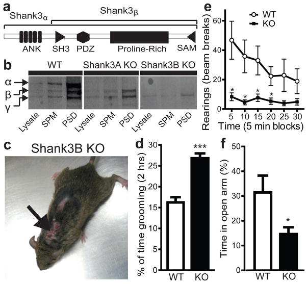
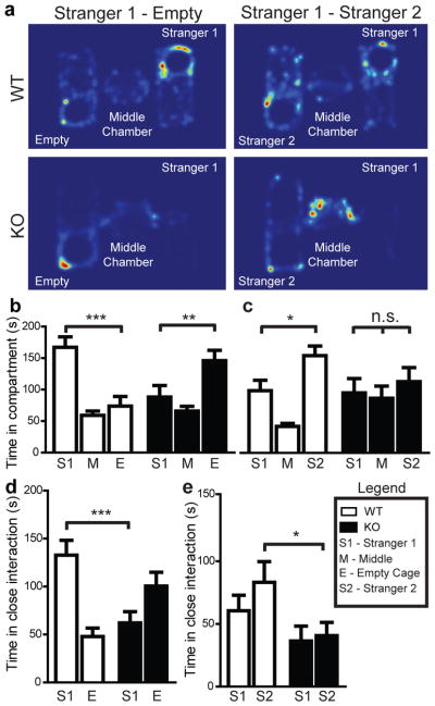
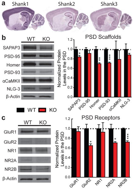
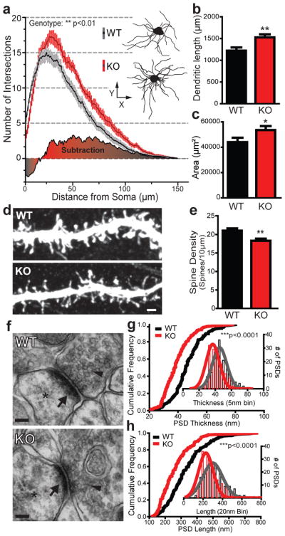
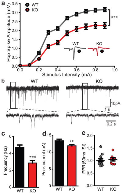
Autism spectrum disorders (ASDs) comprise a range of disorders that share a core of neurobehavioural deficits characterized by widespread abnormalities in social interactions, deficits in communication as well as restricted interests and repetitive behaviours. The neurological basis and circuitry mechanisms underlying these abnormal behaviours are poorly understood. SHANK3 is a postsynaptic protein, whose disruption at the genetic level is thought to be responsible for the development of 22q13 deletion syndrome (Phelan-McDermid syndrome) and other non-syndromic ASDs. Here we show that mice with Shank3 gene deletions exhibit self-injurious repetitive grooming and deficits in social interaction. Cellular, electrophysiological and biochemical analyses uncovered defects at striatal synapses and cortico-striatal circuits in Shank3 mutant mice. Our findings demonstrate a critical role for SHANK3 in the normal development of neuronal connectivity and establish causality between a disruption in the Shank3 gene and the genesis of autistic-like behaviours in mice.


Conflict of interest statement

Author Information: The authors declare no competing financial interests.

Figures

Figure 1
Figure 1. Excessive grooming, skin lesions and anxiety-like behaviour in Shank3B/ mice
a, Shank3 protein structure. b, Western blot showing a pan-Shank3 antibody staining in brain lysate, synaptosomal plasma membrane (SPM) and PSD 2T fraction in wildtype (WT), Shank3A/ and Shank3B/ mice. c, Four month old Shank3B/ mice display neck and head lesions (arrows). d, Pre-lesion Shank3B/ (KO) mice spent more time in self-grooming than WT. e, In the open field test, Shank3B/ mice, when compared to controls, display decreased rearing activity. f, In the zero maze test, Shank3B/ mice spent less time in the open area than wildtype controls. * p<0.05, *** p<0.001, two-tailed t-test for d and f, Two-way repeated measures ANOVA with post hoc two-tailed t-test for e; all data are presented as means ± s.e.m. from 6–9 mice per genotype.
Figure 2
Figure 2. Reduced social interaction and abnormal social novelty recognition in Shank3B/mice
a, Representative heat map analysis from “Stranger 1 Empty” and “Stranger 1 Stranger 2” trials from Shank3B/ mice and controls. b, In the social interaction test, Shank3B/ mice (closed bars) spent less time in the chamber containing the social partner (Stranger 1) and more time in the chamber containing the empty wire cage (Empty Cage) when compared to controls (open bars). c, In the social novelty test, Shank3B/ mice do not display a preference for the novel social partner (Stranger 2), and spent more time in the middle chamber. d, e, When analyzing social interaction by close proximity (within 5 cm) to either “Stranger 1”, “Empty Cage” (d), or “Stranger 1”, “Stranger 2” (e), Shank3B/ mice displayed a clear reduction in social interaction when compared to controls (d); while under a social novelty paradigm (e), Shank3B/ mice displayed a clear reduction in time spent with “Stranger 2 * p<0.05, ** p<0.01, *** p<0.0001; One-way ANOVA, with Bonferroni post hoc t-test for be; all data presented as means ± s.e.m; 12–14 mice per group.
Figure 3
Figure 3. Biochemical changes in striatal synapses of Shank3B/ mice
a, Only Shank3 mRNA is highly expressed in the striatum. b, Protein levels of the scaffolding proteins SAPAP3, Homer and PSD93 are reduced in striatal PSD fractions from Shank3B/ mice. c, Protein levels of glutamate receptor subunits GluR2, NR2A and NR2B are reduced in striatal PSD fractions from Shank3B/ mice. Each lane was loaded with 3 μg of protein with β-Actin as loading control and normalized to wildtype levels. * p<0.05, ** p<0.01, *** p<0.001, two-tailed t-test; all data are presented as means ± s.e.m; n=3 samples per group, with each sample being a combined pool of striatal tissue from 3 animals.
Figure 4
Figure 4. Morphological and ultrastructural neuronal abnormalities in Shank3B/ mice
a, Sholl analysis reveals an increased neuronal complexity of Shank3B/ MSNs (red) when compared to MSNs from wildtype mice (grey); example neurons are shown as insets (WT top; KO bottom). b, c, MSNs from Shank3B/ mice show an increase in total dendritic length (b) and surface area (c) when compared to controls. d, Representative confocal stacks of dye-filled MSNs from KO and WT mice; scale bar 1μm. e, Spine density in MSNs from Shank3B/ mice is lower than that of wildtype MSNs. f, Examples of electron micrographs depicting the synaptic contacts with presynaptic vesicles (arrowheads), postsynaptic densities (arrow) and dendritic spine (asterisk); scale bar 100 nm. g, Shank3B/ PSDs are thinner than wildtype PSDs. h, Shank3B/ PSDs are shorter than wildtype PSDs. * p<0.05, ** p<0.01, *** p<0.0001; Two-Way repeated measures ANOVA for a; two-tailed t-test for b, c and e; Two-sample Kolmogorov-Smirnov test for g and h. Data in g and h are presented as cumulative frequency plot with histogram distribution and Gaussian curve fit for the insets. Data from b, c and e are presented as means ± s.e.m; n= 36 from 3 wildtype mice and n= 36 from 3 Shank3B/ mice for ac; n=41 dendritic segments from 3 wildtype mice and n=36 dendritic segments from 3 Shank3B/ mice for e; n=144 PSDs from 3 wildtype mice and n= 140 PSDs from 3 Shank3B/ mice g, h.
Figure 5
Figure 5. Reduced cortico-striatal synaptic transmission in Shank3B/ MSNs
a, Cortico-striatal pop spike amplitude is decreased in Shank3B/ mice (red trace) as measured by extracellular field recordings. Inset, example traces for Shank3B/ (KO) and wildtype (WT). b, mEPSC example traces from wildtype and Shank3B/ MSNs recorded with whole-cell voltage clamp. c, d, Reduced mEPSC frequency (c) and amplitude (d) in Shank3B/ MSNs when compared to wildtype. e, PPR is unaltered in Shank3B/ MSNs. ** p<0.01, *** p<0.001; Two-way repeated measures ANOVA, with Bonferroni post hoc test for a; two-tailed t-test for c, d; all data presented as means ± s.e.m. For field recordings, n=13 slices from 4 mice per group; for mEPSCs, n=29 MSNs from wildtype mice, n=32 MSNs from Shank3B/ mice.

Comment in

References

    1. American Psychiatric Association. Diagnostic and statistical manual of mental disorders: DSM-IV-TR. American Psychiatric Association; Washington, DC: 2000. Task Force on DSM-IV.
    1. Rosenberg RE, et al. Characteristics and concordance of autism spectrum disorders among 277 twin pairs. Arch Pediatr Adolesc Med. 2009;163:907–914. - PubMed
    1. Abrahams BS, Geschwind DH. Advances in autism genetics: on the threshold of a new neurobiology. Nat Rev Genet. 2008;9:341–355. - PMC - PubMed
    1. Bourgeron T. A synaptic trek to autism. Current Opinion in Neurobiology. 2009;19:231–234. - PubMed
    1. Zoghbi HY. Postnatal neurodevelopmental disorders: meeting at the synapse? Science. 2003;302:826–830. - PubMed

Publication types

MeSH terms