. 2011 Apr 21;472(7343):325-30.
doi: 10.1038/nature09853. Epub 2011 Mar 20.

Crystal structure of inhibitor of κB kinase β

Affiliations

Crystal structure of inhibitor of κB kinase β

Guozhou Xu et al. Nature. .

Abstract

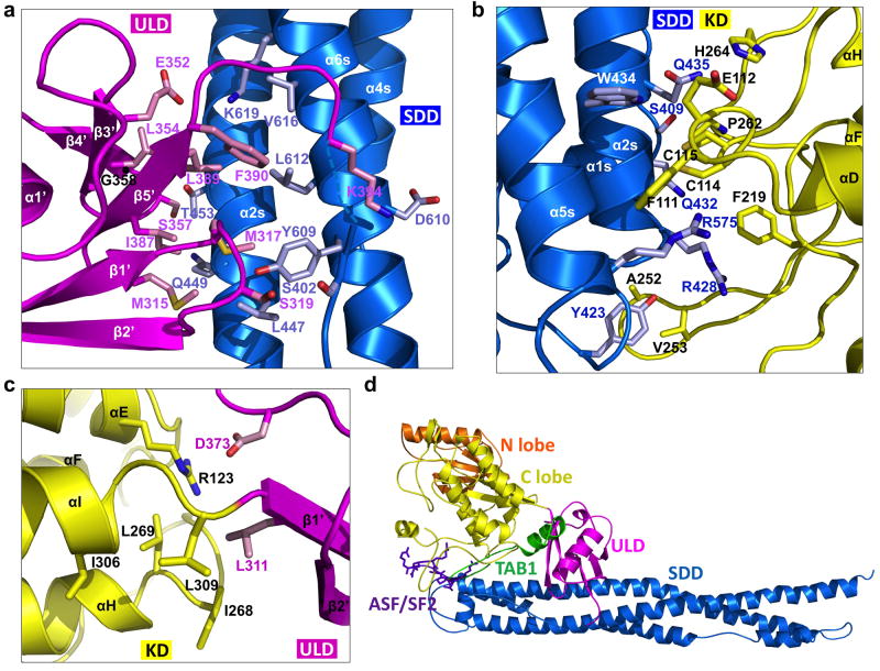
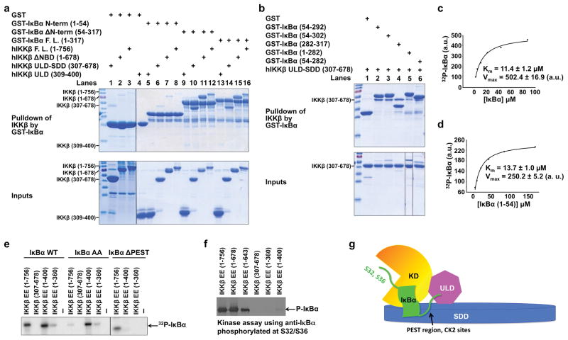
Inhibitor of κB (IκB) kinase (IKK) phosphorylates IκB proteins, leading to their degradation and the liberation of nuclear factor κB for gene transcription. Here we report the crystal structure of IKKβ in complex with an inhibitor, at a resolution of 3.6 Å. The structure reveals a trimodular architecture comprising the kinase domain, a ubiquitin-like domain (ULD) and an elongated, α-helical scaffold/dimerization domain (SDD). Unexpectedly, the predicted leucine zipper and helix-loop-helix motifs do not form these structures but are part of the SDD. The ULD and SDD mediate a critical interaction with IκBα that restricts substrate specificity, and the ULD is also required for catalytic activity. The SDD mediates IKKβ dimerization, but dimerization per se is not important for maintaining IKKβ activity and instead is required for IKKβ activation. Other IKK family members, IKKα, TBK1 and IKK-i, may have a similar trimodular architecture and function.


Figures

Figure 1
Figure 1. Structure of xIKKβ
a, Linear representation of IKKβ showing the boundaries for the kinase domain (KD), the ubiquitin-like domain (ULD) and the scaffold/dimerization domain (SDD). Sequences of hIKKβ and xIKKβ are of 756 and 758 residues, respectively, differing only at the most C-terminal region. The crystallized xIKKβ construct is shown. The previously designated leucine zipper (LZ) and helix-loop-helix (HLH) regions are shown in parentheses. b, Ribbon diagram of an xIKKβ protomer in the P1 crystal form. The N- and C-termini, KD N-lobe (orange) and C-lobe (yellow), ULD (magenta) and SDD (blue) are labeled. Secondary structural elements are labeled, with those in ULD followed by (‘) and those in SDD followed by (s). c, Ribbon diagram of an xIKKβ dimer. d, Superposition of ULD (magenta) with ubiquitin (gray). e, Ribbon diagram of an SDD dimer, showing locations of the previously designated LZ (red) and HLH (orange) regions.
Figure 2
Figure 2. Inhibitor bound xIKKβ kinase domain (KD)
a, Fo-Fc electron density map for Cmpd1 in the I4122 structure, contoured at 2.0 σ. Carbon, nitrogen and oxygen atoms are shown in green, blue and red, respectively. b, Structure of xIKKβ KD. Glycine-rich loop: cyan; activation segment: red except that the DLG and APE motifs are in black; Cmpd1: purple. Side chains of phosphomimic residues E177 and E181 are shown. c, Superposition between xIKKβ (orange and yellow) and PKA (cyan, PDB code 1ATP). The activation segments of xIKKβ and PKA are shown in red and black, respectively.
Figure 3
Figure 3. Interactions among the KD, the ULD and the SDD
a, Interaction between ULD (magenta) and SDD (blue). Important interfacial side chains are shown with nitrogen atoms in blue, oxygen atoms in red, sulfur atoms in orange, and carbon atoms in either pink (for ULD) or light blue (for SDD). Location of G358 is marked with a black ball on the main chain. b, Interaction between KD (yellow) and SDD (blue). c, Interaction between KD (yellow) and ULD (magenta). d, Locations of the TAB1 peptide (green ribbon, PDB code 2EVA) and the ASF/SF2 peptide (purple stick model, PDB code 1WBP) relative to the IKKβ structure after superposition of the corresponding kinase domains.
Figure 4
Figure 4. ULD-SDD restricts IKKβ specificity while ULD is required for catalytic activity
a, b,Pulldown of hIKKβ constructs using GST-IκBα constructs, showing the reciprocal interaction between ULD-SDD of IKKβ and C-terminal region of IκBα containing ankyrin repeats and PEST region. c, d, Measurement of Km and relative Vmax of IKKβ against full-length IκBα (c) and the N-terminal region of IκBα (1–54) (d). a.u.: arbitrary unit. e, Kinase assay of purified hIKKβ proteins against IκBα, its S32A/S36A mutant (AA), or its PEST-deletion construct (ΔPEST, 1–282) using [γ-32P]ATP. f, Kinase assay of purified hIKKβ proteins using antibody against IκBα phosphorylated at S32 and S36. g, A schematic model showing that the interaction between SDD of IKKβ and C-terminal region of IκBα may position the N-terminal cognate phosphorylation sites of IκBα to the active site of IKKβ.
Figure 5
Figure 5. Dimerization is critical for IKKβ activation but not for its activity
a, Gel filtration profiles of various hIKKβ constructs showing that those containing the KD-ULD-SDD region (1–756, 1–678) are dimeric while a further truncated construct (1–643) is monomeric. b, The dimerization interface of xIKKβ. c, Structure-based mutations disrupt hIKKβ dimerization as shown by gel filtration and analytical ultracentrifugation. d, Kinase activity of HEK293T cell transfected or insect cell purified hIKKβ EE and dimerization defective mutants L654D/W655D (654–5), W655D/L658D (655–8) and L654D/W655D/L658D (654-5-8). e, Autoactivation of HEK293T cell transfected hIKKβ WT and its dimerization defective mutants. f, Transfection of hIKKβ into WT and NEMO−/− MEFs, showing reduced IKKβ activation in the absence of NEMO. g, Dimerization mutants of IKKβ showed reduced interaction with NEMO. h, A tetramer of xIKKβ in the P1 structure. i, A close-up view of the tetramer interface, showing that the activation loops of neighboring protomers (black) face each other.

Comment in

References

    1. Hayden MS, Ghosh S. Shared principles in NF-kappaB signaling. Cell. 2008;132 (3):344. - PubMed
    1. Vallabhapurapu S, Karin M. Regulation and function of NF-kappaB transcription factors in the immune system. Annu Rev Immunol. 2009;27:693. - PubMed
    1. Scheidereit C. IkappaB kinase complexes: gateways to NF-kappaB activation and transcription. Oncogene. 2006;25 (51):6685. - PubMed
    1. Karin M. Nuclear factor-kappaB in cancer development and progression. Nature. 2006;441 (7092):431. - PubMed
    1. Chen ZJ, Parent L, Maniatis T. Site-specific phosphorylation of IkappaBalpha by a novel ubiquitination-dependent protein kinase activity. Cell. 1996;84 (6):853. - PubMed

Publication types