. 2011 Jul 21;30(29):3207-21.
doi: 10.1038/onc.2011.35. Epub 2011 Mar 21.

The pro-longevity gene FoxO3 is a direct target of the p53 tumor suppressor

Affiliations

The pro-longevity gene FoxO3 is a direct target of the p53 tumor suppressor

V M Renault et al. Oncogene. .

Abstract

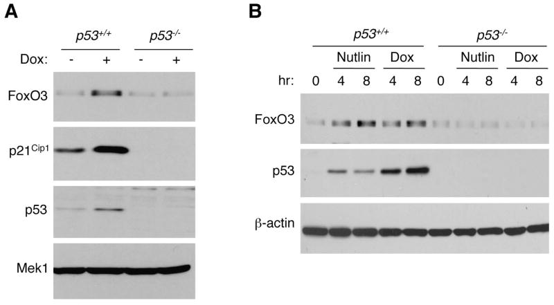
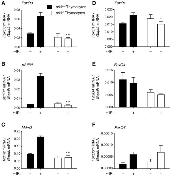
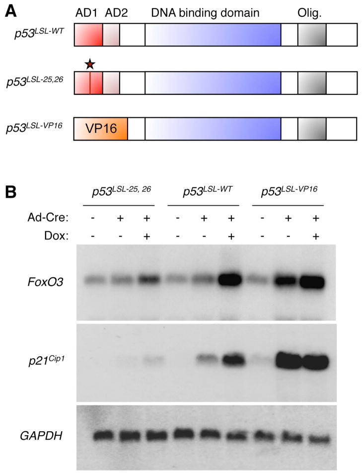
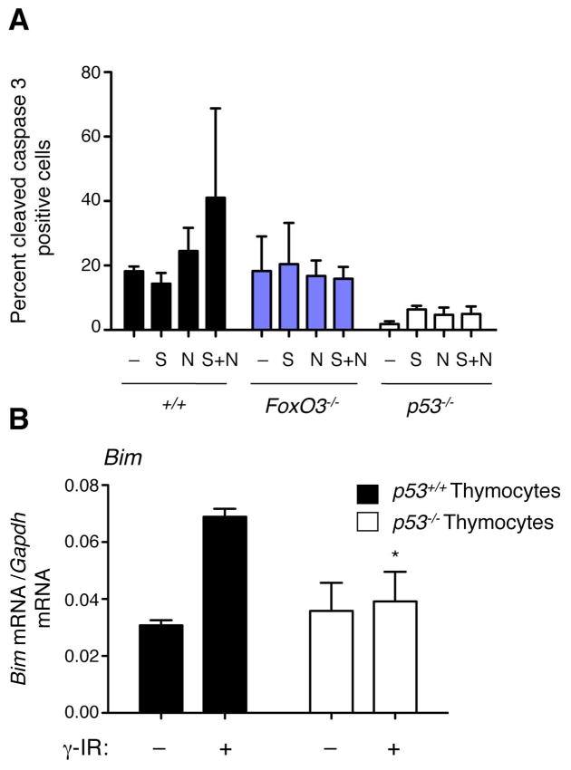
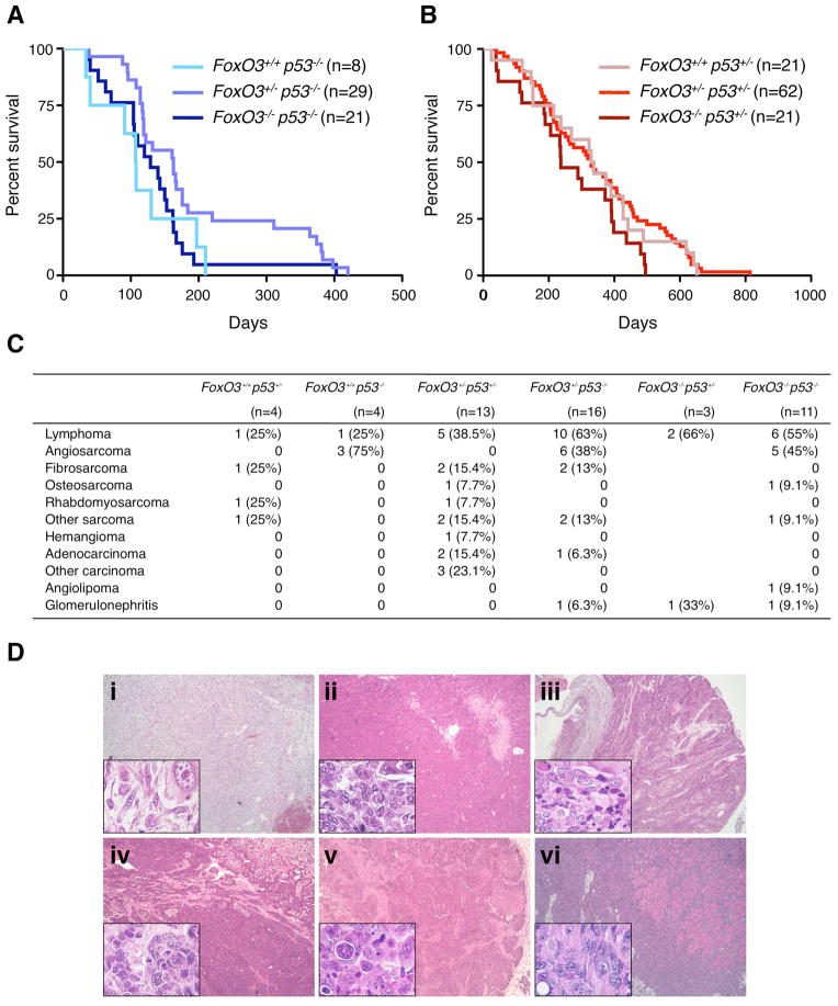
FoxO transcription factors have a conserved role in longevity, and act as tissue-specific tumor suppressors in mammals. Several nodes of interaction have been identified between FoxO transcription factors and p53, a major tumor suppressor in humans and mice. However, the extent and importance of the functional interaction between FoxO and p53 have not been fully explored. Here, we show that p53 regulates the expression of FoxO3, one of the four mammalian FoxO genes, in response to DNA damaging agents in both mouse embryonic fibroblasts and thymocytes. We find that p53 transactivates FoxO3 in cells by binding to a site in the second intron of the FoxO3 gene, a genomic region recently found to be associated with extreme longevity in humans. While FoxO3 is not necessary for p53-dependent cell cycle arrest, FoxO3 appears to modulate p53-dependent apoptosis. We also find that FoxO3 loss does not interact with p53 loss for tumor development in vivo, although the tumor spectrum of p53-deficient mice appears to be affected by FoxO3 loss. Our findings indicate that FoxO3 is a p53 target gene, and suggest that FoxO3 and p53 are part of a regulatory transcriptional network that may have an important role during aging and cancer.


Conflict of interest statement

Conflict of interest

We declare that there are no competing financial interests in relation to the work described.

Figures

Figure 1
Figure 1. Doxorubicin and Nutlin elicit an increase in FoxO3 protein expression that is p53-dependent in MEFs
(A) Western-blot of protein extracts from p53+/+ and p53−/− MEFs incubated in the absence (−) or presence (+) of doxorubicin (Dox, 0.2 μg/ml) for 8 hours, using antibodies to FoxO3, p21Cip1 (a well-known target of p53), p53, and Mek1 (loading control). (B) Western-blot of protein extracts from p53+/+ and p53−/− MEFs incubated with Nutlin (10 μM), a p53 activator, or Doxorubicin (Dox, 0.2 μg/ml) for 0, 4, and 8 hours, using antibodies to FoxO3, p53, and β-actin (loading control). Western-blots are representative of at least two independent experiments, conducted on independent cultures of MEFs.
Figure 2
Figure 2. p53 is necessary for FoxO3 mRNA upregulation in response to doxorubicin or Nutlin in MEFs
Real time quantitative PCR analysis of FoxO3 (A), p21Cip1 (B), Mdm2 (C), FoxO1 (D), FoxO4 (E), and FoxO6 (F) mRNA levels in p53+/+ and p53−/− MEFs in response to 4 and 8 hours of treatment with Nutlin (10 μM) or Doxorubicin (Dox, 0.2 μg/ml). Mean +/− SEM of two independent experiments conducted in triplicate. * p<0.05, ** p<0.01, ***p<0.001 between p53+/+ and p53−/− MEFs at a given time point, two-way ANOVA with Bonferroni post-test.
Figure 3
Figure 3. p53 is necessary for FoxO3 mRNA upregulation in response to γ irradiation in thymocytes
Real time quantitative PCR analysis of FoxO3 (A), p21Cip1 (B), Mdm2 (C), FoxO1 (D), FoxO4 (E), and FoxO6 (F) mRNA levels in p53+/+ and p53−/− thymocytes, 3 hours after γ irradiation (γ IR, 10 Gy). Mean +/− SEM of two independent experiments conducted in triplicate on samples from 3–5 mice per genotype. * p<0.05, ***p<0.001 between p53+/+ and p53−/− thymocytes at a given time point, two-way ANOVA with Bonferroni post-test.
Figure 4
Figure 4. p53 transcriptional activity is necessary and sufficient for FoxO3 mRNA upregulation
(A) Schematic of the p53 knock-in alleles used. p53LSL-WT: inducible allele encoding a form of wildtype p53. p53LSL-25,26: inducible allele encoding a transcriptionally-impaired mutant of p53 in which leucine 25 is replaced by a glutamine and tryptophan 26 is replaced by a serine. p53LSL-VP16: inducible allele encoding a mutant of p53 in which the transactivation domains (AD1 and AD2) are replaced by the transactivation domain of VP16. AD: activation domain; Olig.: oligomerization domain. The star indicates the location of the 25,26 mutation. (B) Northern-blot analysis of MEFs in which the endogenous allele of p53 has been replaced by an allele encoding inducible forms of WT p53 (p53LSL-WT), a transcription-deficient mutant (p53LSL-25,26), or a mutant of p53 in which the transactivation domains of p53 were replaced by that of VP16 (p53LSL-VP16). The addition of an adenovirus containing Cre recombinase (Ad-Cre) allows the deletion of the Lox-STOP-Lox (LSL) cassette upstream of each allele and allows the expression of each p53 variant. Cells were exposed to 8 hours of doxorubicin (Dox, 0.2 μg/ml). Northern-blots were analyzed with a probe to FoxO3, p21Cip1 (a known target of p53), and GAPDH (loading control).
Figure 5
Figure 5. p53 is recruited to a binding site in the second intron of the FoxO3 gene
(A) Location and sequence of the p53 binding sites (p53-1, p53-2, p53-3, and p53-4) in the promoter and second intron of the mouse FoxO3 gene. R: G or A; W: T or A; Y: C or T; E: exon; I: intron. Also depicted are the consensus for p53 binding sites, the p53 binding site in p21Cip1 promoter, and the mutant of critical bases in p53-4 (p53-4m). (B) ChIP on MEFs treated with Doxorubicin (Dox, 0.2 μg/ml) for 16–20 hours, using antibodies to p53 (colored bars) or control IgG (white bars). The chromatin bound to p53 or to the control IgG was analyzed by quantitative-PCR with primers surrounding a region that did not contain p53 binding sites (−), the distal p53 binding site in FoxO3 promoter (p53-1), the p53-4 binding site in FoxO3 intron 2 (p53-4), and the p53 binding site in the p21Cip1 promoter (p53 p21Cip1). The fold enrichment over the IgG control is represented. Mean +/− SEM of three independent experiments. **: p<0.01, one-way ANOVA. (C) ChIP on p53+/+ and p53−/− MEFs in the absence or presence of Doxorubicin (Dox, 0.2 μg/ml) for 6 hours, using antibodies to p53 or control IgG. The chromatin bound to p53 or to the control IgG was analyzed by quantitative-PCR with primers surrounding a region that did not contain p53 binding sites (−), the distal p53 binding site in FoxO3 promoter (p53-1), the p53-4 binding site in FoxO3 intron 2 (p53-4) and the p53 binding site in the p21Cip1 promoter (p53 p21Cip1). The fold enrichment over the IgG control is represented. Mean +/− SD from triplicates of one experiment. (D) Normalized activity of luciferase reporter constructs driven by 500bp surrounding the p53 binding sites p53-1 or p53-4 in p53+/+ (black) and p53−/− (white) MEFs. Mean +/− SEM of four independent experiments conducted in triplicate. *: p<0.05 between p53-4 and control in p53+/+ MEFs, **: p<0.01 between p53+/+ and p53−/− MEFs for p53-4, one-way ANOVA. (E) Normalized activity of luciferase reporter constructs driven by the region surrounding the p53-4 binding site or by the region surrounding the p53-4 binding site in which the p53 binding site was mutated (p53-4m) in p53+/+ (black) and p53−/− (white) MEFs. Mean +/− SEM of three independent experiments conducted in triplicate. **: p<0.01 between p53-4m and p53-4 in p53+/+ MEFs, one-way ANOVA.
Figure 6
Figure 6. FoxO3 is not necessary for p53-dependent cell cycle arrest in MEFs, but other FoxO family members may compensate for FoxO3 loss
(A) Percent BrdU-positive cells in FoxO3+/+p53+/+ (+/+), FoxO3−/− and p53−/− MEFs in the presence or absence of doxorubicin (Dox, 0.2 μg/ml) for 24 hours. Mean +/− SEM of three independent experiments, two of which were conducted with independent MEF cultures from distinct animals. ***p<0.001 between +/+ and p53−/− MEFs for the same treatment; ns: non significant between +/+ and FoxO3−/− MEFs for the same treatment, two-way ANOVA with Bonferroni post-test. (B) Percent BrdU-positive cells in FoxO3+/+p53+/+ (+/+), FoxO3−/− and p53−/− MEFs in the presence or absence of Nutlin (10 μM) for 24-36 hours. Mean +/− SEM of three independent experiments. *p<0.05, ***p<0.001 between +/+ and p53−/− MEFs for the same treatment; ns: non significant between +/+ and FoxO3−/− MEFs for the same treatment, two-way ANOVA with Bonferroni post-test. (C) Percent BrdU-positive cells in FoxO3+/+p53+/+ (+/+), FoxO3−/− and p53−/− MEFs in the presence or absence of chronic treatment of H2O2 or hydroxyurea (HU). Mean +/− SEM of two independent experiments with different lines of MEFs. **p<0.01, ***p<0.001 between +/+ and FoxO3−/− or p53−/− MEFs for the same treatment; ns: non significant between +/+ and FoxO3−/− MEFs for the same treatment, two-way ANOVA with Bonferroni post-test. (D) Real time quantitative PCR analysis of p27Kip1 mRNA levels in p53+/+ and p53−/− MEFs in response to 4 and 8 hours of treatment with Nutlin (10 μM) or Doxorubicin (Dox, 0.2 μg/ml). Mean +/− SEM of two independent experiments conducted in triplicate. ***p<0.001 between p53+/+ and p53−/− MEFs at a given time point, two-way ANOVA with Bonferroni post-test. (E) Percent BrdU-positive cells in MEFs infected with control lentiviruses (PSR) or with lentiviruses expressing an shRNA to FoxO family members (PSR “pan FoxO”) in the presence or absence of Nutlin (Nutlin, 10 μM) for 24 hours. The data are expressed as fold decrease, respective to the value obtained in the absence of Nutlin. Mean +/− SEM of two independent experiments. ** p<0.01, between PSR and PSR “pan” FoxO-infected MEFs in the presence of Nutlin, two-way ANOVA with Bonferroni post-test. (F) Western-blot of protein extracts from MEFs infected with control lentiviruses (PSR) or with lentiviruses expressing an shRNA to FoxO family members (PSR “pan FoxO”), using antibodies to FoxO1, FoxO3, FoxO4, FoxO6, and β actin.
Figure 7
Figure 7. FoxO3 plays a role in p53-dependent apoptosis
(A) Percent cleaved caspase 3-positive cells in E1A-transformed MEFs (FoxO3+/+p53+/+ (+/+), FoxO3−/−, and p53−/−) in the absence of treatment (−) or in response to Nutlin (N), serum starvation (S), and Nutlin + serum starvation (N+S). Mean +/− SEM of three independent experiments. (B) Real time quantitative PCR analysis of Bim mRNA levels in p53+/+ and p53−/− thymocytes, 3 hours after γ irradiation (γ IR, 10 Gy). Mean +/− SEM of two independent experiments conducted in triplicate on samples from 3–5 mice per genotype. * p<0.05 between p53+/+ and p53−/− thymocytes at a given time point, two-way ANOVA with Bonferroni post-test.
Figure 8
Figure 8. FoxO3 loss does not affect survival in mice that are lacking one or both p53 alleles, but may alter tumor spectrum
(A). Percent survival of mice with different alleles of FoxO3 in the p53−/− background as a function of time. Kaplan-Meier survival curves with the number of mice indicated for each genotype. p = 0.10, logrank test. (B) Percent survival of mice with different alleles of FoxO3 in the p53+/− background as a function of time. Kaplan-Meier survival curves with the number of mice indicated for each genotype. p = 0.13, logrank test. (C) Tumor types and glomerulonephritis in mice with different alleles of FoxO3 in the p53+/− and p53−/− background. The number of mice is indicated for each genotype. (D) Examples of sarcomas and carcinomas in compound FoxO3/p53 mutant mice. Main panels: 50x. Insets: 630x. i: subcutaneous fibrosarcoma in a FoxO3+/−p53−/− mouse; ii: osteosarcoma in the leg of a FoxO3−/−p53−/− mouse; iii: colon carcinoma in a FoxO3+/−p53−/− mouse; iv: uterine carcinoma in a FoxO3+/−p53+/− mouse; v: breast carcinoma in a FoxO3+/−p53+/− mouse; vi: muscle carcinoma in the arm of a FoxO3+/−p53+/− mouse.

Similar articles

Cited by

References

    1. Anselmi CV, Malovini A, Roncarati R, Novelli V, Villa F, Condorelli G, et al. Association of the FOXO3A locus with extreme longevity in a southern Italian centenarian study. Rejuvenation Res. 2009;12:95–104. - PubMed
    1. Arum O, Johnson TE. Reduced expression of the Caenorhabditis elegans p53 ortholog cep-1 results in increased longevity. J Gerontol A Biol Sci Med Sci. 2007;62:951–9. - PubMed
    1. Attardi LD, Reczek EE, Cosmas C, Demicco EG, McCurrach ME, Lowe SW, et al. PERP, an apoptosis-associated target of p53, is a novel member of the PMP-22/gas3 family. Genes Dev. 2000;14:704–18. - PMC - PubMed
    1. Bakker WJ, Harris IS, Mak TW. FOXO3a is activated in response to hypoxic stress and inhibits HIF1-induced apoptosis via regulation of CITED2. Mol Cell. 2007;28:941–53. - PubMed
    1. Bauer JH, Poon PC, Glatt-Deeley H, Abrams JM, Helfand SL. Neuronal expression of p53 dominant-negative proteins in adult Drosophila melanogaster extends life span. Curr Biol. 2005;15:2063–8. - PubMed

Publication types

MeSH terms