. 2011 Feb 17;6(2):e16992.
doi: 10.1371/journal.pone.0016992.

PTBP1 is required for embryonic development before gastrulation

Affiliations

PTBP1 is required for embryonic development before gastrulation

Jakob Suckale et al. PLoS One. .

Abstract

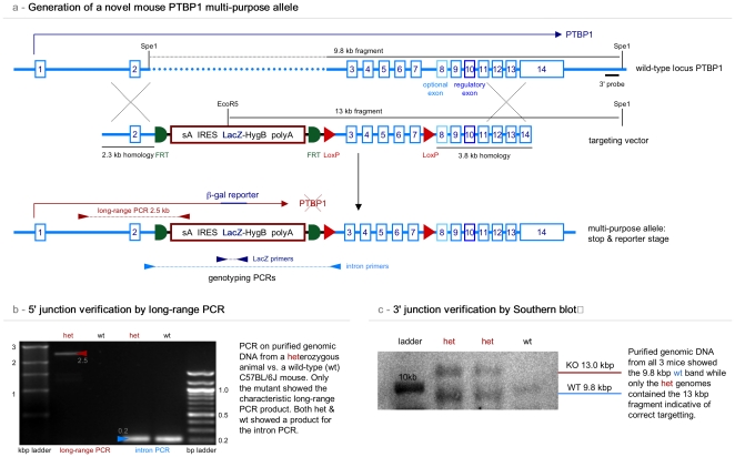
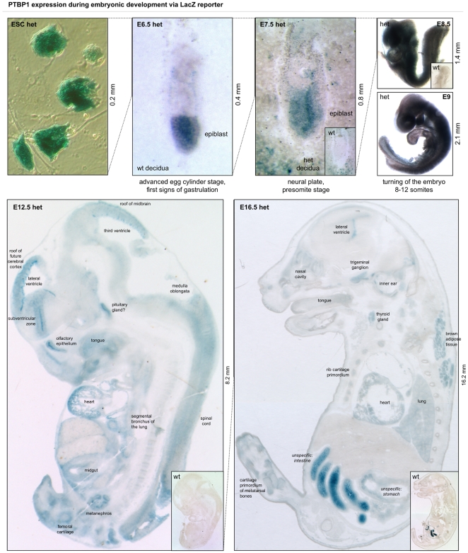
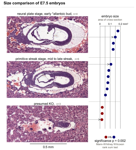
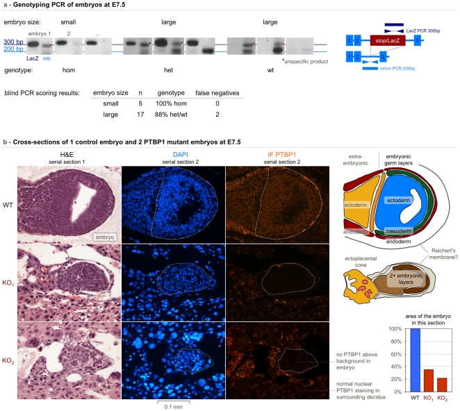
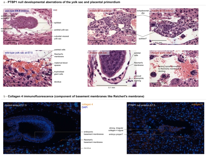
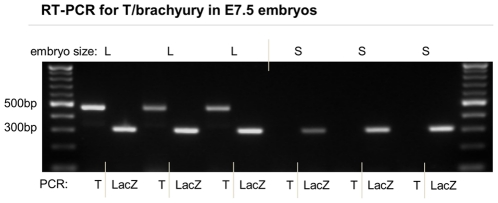
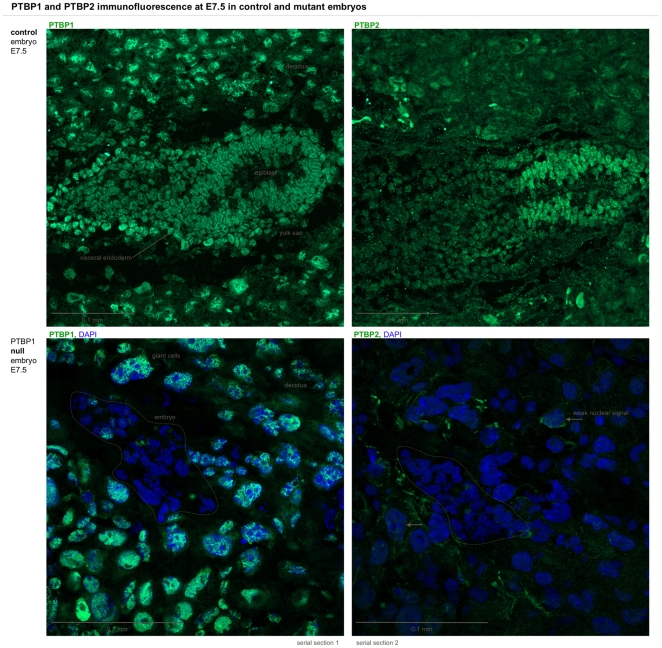
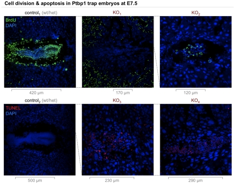
Polypyrimidine-tract binding protein 1 (PTBP1) is an important cellular regulator of messenger RNAs influencing the alternative splicing profile of a cell as well as its mRNA stability, location and translation. In addition, it is diverted by some viruses to facilitate their replication. Here, we used a novel PTBP1 knockout mouse to analyse the tissue expression pattern of PTBP1 as well as the effect of its complete removal during development. We found evidence of strong PTBP1 expression in embryonic stem cells and throughout embryonic development, especially in the developing brain and spinal cord, the olfactory and auditory systems, the heart, the liver, the kidney, the brown fat and cartilage primordia. This widespread distribution points towards a role of PTBP1 during embryonic development. Homozygous offspring, identified by PCR and immunofluorescence, were able to implant but were arrested or retarded in growth. At day 7.5 of embryonic development (E7.5) the null mutants were about 5x smaller than the control littermates and the gap in body size widened with time. At mid-gestation, all homozygous embryos were resorbed/degraded. No homozygous mice were genotyped at E12 and the age of weaning. Embryos lacking PTBP1 did not display differentiation into the 3 germ layers and cavitation of the epiblast, which are hallmarks of gastrulation. In addition, homozygous mutants displayed malformed ectoplacental cones and yolk sacs, both early supportive structure of the embryo proper. We conclude that PTBP1 is not required for the earliest isovolumetric divisions and differentiation steps of the zygote up to the formation of the blastocyst. However, further post-implantation development requires PTBP1 and stalls in homozygous null animals with a phenotype of dramatically reduced size and aberration in embryonic and extra-embryonic structures.


Conflict of interest statement

Competing Interests: The authors have declared that no competing interests exist.

Figures

Figure 1
Figure 1. Generation of a mouse containing a novel multipurpose PTBP1 allele.
We generated a multipurpose PTBP1 allele and targeted it into mice. A) The wild-type locus (blue) was modified with the insertion of a stop/detection cassette (red) into embryonic stem cells. The cassette can be flipped out using an FLP recombinase construct or strain. In addition, exons 3 to 7 have been framed with loxP sites for conditional removal by a Cre recombinase. The diagram indicates the Southern strategy as well as the location of the primers for validation of the construct and genotyping. B) shows the results of the long-range PCR to verify the 5′ junction resulting from the homologous recombination of the targeting vector with the wild-type PTBP1 locus. C) Southern blot to verify the correct 3′ junction in the mutant allele.
Figure 2
Figure 2. PTBP1 is expressed throughout embryonic development.
The LacZ reporter shown in Figure 1 was used here as a proxy for PTBP1 promoter activity in heterozygous embryonic stem cells (ESCs) and embryos at different stages of development. PTBP1 expression was high in ESCs. In embryos 6.5 days after fertilisation (E6.5), PTBP1 was expressed especially in the epiblast but also in the visceral endoderm surrounding the embryo. The expression pattern was similar at E7.5 where the activation of maternal PTBP1 was seen in the decidua surrounding the tip of the embryo. No LacZ staining was observed in the wild-type (wt) littermate (small inlet). The reporter was expressed in almost all cells at E8.5 and in most cells at E9, when only the areas at the roof of the future head, the primitive ventricle of the heart, and some structures in the tail appeared to express little or no PTBP1. In E12.5 embryos most developing tissues and organs expressed the reporter, most notably the cells lining the ventricles of the brain, the spinal cord, the pituitary and the heart. Similar organs were stained in E16.5 embryos and in addition the nasal cavities, the inner ear and the brown fat pad on the back of the embryo. PTBP1 was high in the ventral rib cartilage but much lower in the dorsal rib bones that already showed a central cavity. While at E12.5 there was no unspecific staining, at E16.5 the intestine and to a lesser degree the stomach displayed an X-gal signal not due to the PTBP1 reporter. The signal result from endogenous β-galactosidase. See Figure S3 for a whole-mount LacZ at E12 and E16.
Figure 3
Figure 3. Embryos from PTBP1 KO heterozygous parents fall into 2 size categories.
Hematoxylin and eosin stained decidual sections of 2 litters were analysed for surface area and structural organisation. Embryos were grouped according to their developmental stage (left) with the furthest developed having reached neural plate stage but most embryos being at the primitive streak stage. The surface area of the embryos in the middle section was measured with the Fiji image software and ranked according to size, which turned out as equivalent to stage (right). Presumed knockouts (red) were significantly smaller than wild-type or heterozygous controls and present in a roughly Mendelian ratio of 3∶10 (ratio 0.3 vs. the expected 0.25). The size of the small embryos might have even been overestimated since it was harder to recognise the embryo in those implantations. We also encountered 2 decidua with no visible embryonic structures.
Figure 4
Figure 4. PTBP1 knockout embryos implant but are severely growth-retarded.
The small category of embryos from a heterozygous intercross were identified by PCR and immunofluorescence as PTBP1 knockout embryos. A) PCR products for the stop cassette and PTBP1 intron 2 were separated on agarose gels and scored blindly. 5/5 small embryos were genotyped as homozygous. 15/17 large control embryos were genotyped as heterozygous or wild-type with 2 false negatives due to the less efficient intron PCR. B) Serial paraffin sections of E7.5 embryos were stained with hematoxylin and eosin (left column) or labelled with DAPI (middle column) and the PTBP1 antibody (right column). The top row shows a large embryo (in an oblique section) with strong nuclear PTBP1 staining of the embryo proper (dashed line) and the surrounding tissue. Both small embryos were characterised by a lack of the nuclear PTBP1 signal while showing nuclear PTBP1 in surrounding, most likely maternal cells. Interpretative diagrams and a quantification of the embryo section area are shown on the right.
Figure 5
Figure 5. PTBP1 null embryos show defects in yolk sac & placenta development.
This figures compares yolk sac and placental structures in knockout and control embryos. A) Hematoxylin and eosin-stained paraffin sections of an E5.5 wild-type embryo, chosen for its comparable size to the null mutants, and an E7.5 yolk sac (left) juxtaposed with mutant E7.5 embryos and mutant yolk sacs, respectively (right). Mutants lack the typical structure of a normal embryo at this stage. If discernable at all they lack a typical yolk sac with a thin Reichert's membrane in close proximity to trophoblast giant cells and the decidua. B) Immunofluorescence for collagen 4 on a section of a control E7.5 embryo is shown next to a similar image from a homozygous embryo. The control embryo shows collagen 4 signal from several basement membranes including the Reichert's membrane of the yolk sac. The null mutant, on the other hand, shows an unusually extended area high in collagen 4, not resembling a membrane. Separate channels for the collagen immunomicroscopy can be seen in Figure S4.
Figure 6
Figure 6. Deletion of PTBP1 leads to the absence of T/brachyury.
The figure compares RT PCR results from normal and mutant embryos for the presence of a gastrulation marker and a control. RNA purified from large (L) and small (S) embryos was used as template for reverse transcriptase (RT) PCR amplification of the primitive streak marker T/brachyury and of a LacZ control to ensure successful RNA purification. None of the small PTBP1 KO homozygous embryos showed the presence of T mRNA while positive for the control RT PCR.
Figure 7
Figure 7. PTBP1 expression in normal and mutant embryos.
Comparison of immunofluorescence from PTBP1 and its paralogue PTBP2 in control and PTBP1 KO embryos at E7.5. The top row shows serial cryosections of a normally sized control embryo stained for PTBP1 (left) and PTBP2 (right). PTBP1 was expressed in most embryonic cells, notably the visceral endoderm and the epiblast, comparable to the LacZ reporter staining in Figure 2. A strong PTBP1 signal was also observed in decidual nuclei. PTBP2 in controls was mostly expressed in the epiblast and absent from most decidual nuclei. PTBP1 null embryos (bottom) showed no PTBP1 or PTBP2 signal, displayed here merged with the DAPI signal to reveal the embryo (dashed line). Again most cells surrounding the embryo displayed a nuclear PTBP1 signal while only very few showed a weak nuclear PTBP2 signal (arrow).
Figure 8
Figure 8. Cell division & apoptosis in mutant embryos.
The figure shows control and Ptbp1 null embryo sections at E7.5 immunolabelled for BrdU (top row) and processed by TUNEL (bottom row). The former is a cell division assay measuring the integration of the nucleotide analogue bromodeoxyuridine (BrdU). The latter is an apoptosis assay based on terminal deoxynucleotidyl transferase dUTP nick end labelling (TUNEL). Control embryos (1st column) showed strong BrdU signal especially in the epiblast, the yolk sac area, and the ectoplacental cone while being negative for TUNEL. Heterozygous decidua were typically BrdU-labelled most strongly in the periphery, visible only in KO1 and cropped in control1 and KO2. Homozygous mutant embryos showed less (KO1) to no BrdU labelling (KO2) varying with embryo and section. Most null mutants analysed showed some degree of TUNEL (exemplified by KO3 and KO4). The longest diameter of the embryo is indicated below each panel and the relative sizes of the panels are indicated with dashed lines.

Similar articles

Cited by

References

    1. Wang H, Dey SK. Roadmap to embryo implantation: clues from mouse models. Nat Rev Genet. 2006;7:185–199. - PubMed
    1. Tam PPL, Loebel DAF. Gene function in mouse embryogenesis: get set for gastrulation. Nat Rev Genet. 2007;8:368–381. - PubMed
    1. Cross JC. How to make a placenta: mechanisms of trophoblast cell differentiation in mice–a review. Placenta. 2005;26(Suppl A):S3–9. - PubMed
    1. Wollerton MC, Gooding C, Wagner EJ, Garcia-Blanco MA, Smith CWJ. Autoregulation of polypyrimidine tract binding protein by alternative splicing leading to nonsense-mediated decay. Mol Cell. 2004;13:91–100. - PubMed
    1. Boutz PL, Stoilov P, Li Q, Lin C, Chawla G, et al. A post-transcriptional regulatory switch in polypyrimidine tract-binding proteins reprograms alternative splicing in developing neurons. Genes Dev. 2007;21:1636–1652. - PMC - PubMed

Publication types

MeSH terms

Substances