. 2010 Jun 10:2:7.
doi: 10.3389/fnsyn.2010.00007. eCollection 2010.

In vivo spike-timing-dependent plasticity in the optic tectum of Xenopus laevis

Affiliations

In vivo spike-timing-dependent plasticity in the optic tectum of Xenopus laevis

Blake A Richards et al. Front Synaptic Neurosci. .

Abstract

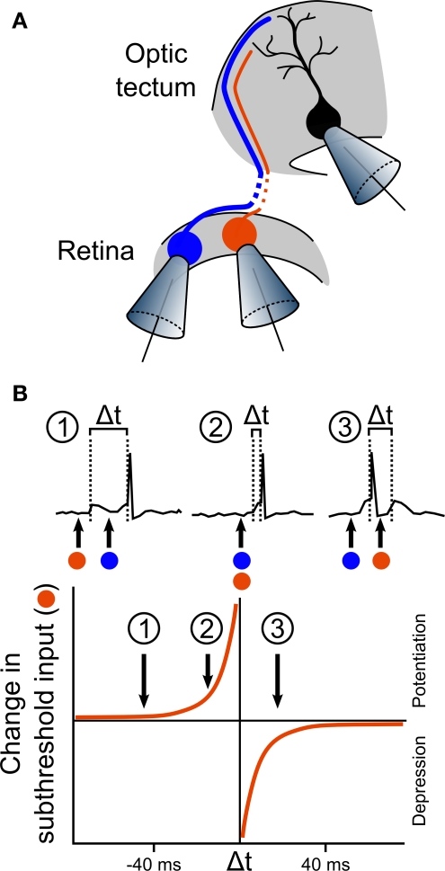
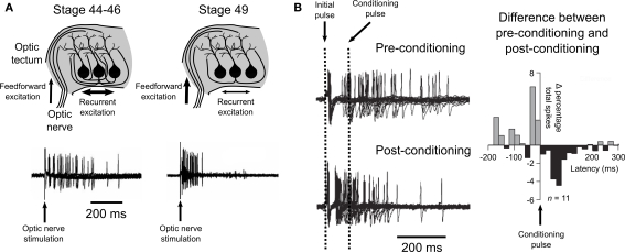
Spike-timing-dependent plasticity (STDP) is found in vivo in a variety of systems and species, but the first demonstrations of in vivo STDP were carried out in the optic tectum of Xenopus laevis embryos. Since then, the optic tectum has served as an excellent experimental model for studying STDP in sensory systems, allowing researchers to probe the developmental consequences of this form of synaptic plasticity during early development. In this review, we will describe what is known about the role of STDP in shaping feed-forward and recurrent circuits in the optic tectum with a focus on the functional implications for vision. We will discuss both the similarities and differences between the optic tectum and mammalian sensory systems that are relevant to STDP. Finally, we will highlight the unique properties of the embryonic tectum that make it an important system for researchers who are interested in how STDP contributes to activity-dependent development of sensory computations.

Keywords: optic tectum; receptive field; spike-timing-dependent plasticity; synaptic development; visual system.


Figures

Figure 1
Figure 1
Anatomy of the optic tectum in the adult Xenopus laevis and during tadpole development. (A) In the adult Xenopus laevis frog (photograph) the central nervous system (drawing) contains several distinct structures and the optic tectum is the roof of the large midbrain structure, situated caudally to the diencephalon and rostrally to the cerebellum. It is one of the largest dorsal structures in the Xenopus brain, along with the telencephalon and the olfactory bulb. (B) The layered structure of the adult optic tectum can be seen in the coronal section (left), which has been stained with cresyl-violet. The section is taken from the plane indicated by the red dashed line in (A). As the drawing illustrates (right), there are several distinct cellular morphologies found within the optic tectum, which have been classified into 14 categories. The numbers at the side of the drawing indicate the 9 different layers of the tectum. (C) Photographs (left) of Xenopus laevis tadpoles taken dorsally at stages 42, 46, and 49 illustrate the changes that occur during the stages in which STDP is typically studied. During this time the optic tectum grows, as shown by confocal images of whole-mount brains with propidium iodide staining for cell nuclei. The images shown are in the horizontal plane and at a depth of 100 μm from the dorsal surface of the brain (right). Note the dark regions in the rostral–lateral optic tectum which are comprised mostly of neuropil. Neurogenesis takes place in the caudal–medial region surrounding the ventricle. (D) Due to the location of the neurogenerative zone there is a progression in the maturity and morphological complexity of cells in the optic tectum at these ages in the caudal–rostral axis, as shown by this camera lucida drawing of a sagittal slice from a stage 49 tadpole. Images in (B) and (D) are reproduced with permission from Lázár (1973) and Nikundiwe and Nieuwenhuys (1983).
Figure 2
Figure 2
First in vivo observation of STDP in the optic tectum. (A) The first in vivo demonstration of STDP was performed by Zhang et al. (1998) in an elegant experiment, illustrated here. Activity from a single tectal neuron was recorded using whole-cell perforated patch, while two different RGCs that form synaptic connections onto the tectal neuron were loose-patched, allowing stimulation for induction of LTP or LTD. In some of the recordings, one RGC produced suprathreshold responses (illustrated by the blue cell), whilst the other produced only subthreshold responses (illustrated by the orange cell). (B) Analysis of the effects of the timing of the inputs showed that the effect of stimulation on the subthreshold input depended on the timing of its excitatory postsynaptic potentials (EPSPs) relative to the tectal spike that was triggered by the suprathreshold input. If the subthreshold EPSP preceded the tectal spike by more than 20 ms, the input was relatively unaffected (inset 1). However, if the subthreshold EPSP occurred within the 20 ms before the suprathreshold EPSP, and therefore just before the tectal spike, the input was strongly potentiated (inset 2). In stark contrast, if the subthreshold EPSP occurred during the 20 ms immediately following the tectal cell spike, this input was depressed (inset 3). Examination of the effects of a range of timing differences led to an estimated curve for the STDP rule in retinotectal synapses.
Figure 3
Figure 3
Development of direction selectivity and RF structure via STDP in the optic tectum. (A) The principle of how STDP can induce direction selectivity in the optic tectum was demonstrated by Mu and Poo (2006), by mimicking movement across the retina with flashes of a white bar at three different locations in visual space. If the tectal cell (black) was forced to spike soon after the second flash, the RF of the cell was altered by STDP in an asymmetric manner that potentiated responses to the first and second bars (green and red cells), but depressed responses to the third bar (blue cell). (B) Asymmetric changes in a RF can produce direction selectivity due to the differences in temporal summation for one direction versus the other. If the strengthened inputs are activated first, they can summate with subsequent inputs to produce a high level of depolarization in postsynaptic tectal cells producing suprathreshold activity (top, dashed line indicates hypothetical spike threshold). In contrast, if the weaker inputs are stimulated first, they will have decayed by the time subsequent inputs arrive and so less temporal summation occurs and inputs remain subthreshold (bottom). (C) The strength of the connections onto a tectal neuron determines its RF profile, as illustrated here for a hypothetical cell. (D) Vislay-Meltzer et al. (2006) demonstrated that this RF profile could be altered by STDP to either move towards or away from a given region of space. If a flash occurred prior to a tectal cell's spikes (red line), the RF tended to shift toward that area. In contrast, if a flash occurred after a tectal cell's spikes (blue line) the RF tended to shift away from the area of the flash. Interestingly, they also observed that the RFs potentiated in areas outside of the area of the flash, as shown.
Figure 4
Figure 4
Spike-timing-dependent plasticity and recurrent excitatory circuits in the optic tectum. (A) Pratt et al. (2008) observed evidence for a developmental refinement of the recurrent excitatory circuitry of the tectum between stages 44 and 49. Stimulation of the optic nerve at early stages (44–46) produced prolonged spiking activity, while stimulation at a later stage (49) produced an initial strong response that did not show prolonged activity, as illustrated by traces from loose-patch recordings shown here. (B) Training young tectum by delivering a timed “conditioning” pulse to the optic nerve induced a similar reshaping of spiking to that observed over development. Examination of the differences in the percentage of spikes that occurred before or after the conditioning pulse showed an effect that supported a role for STDP in this refinement of recurrent circuitry. Data are reproduced from Pratt et al. (2008).

Similar articles

Cited by

References

    1. Aizenman C. D., Akerman C. J., Jensen K. R., Cline H. T. (2003). Visually driven regulation of intrinsic neuronal excitability improves stimulus detection in vivo. Neuron 39, 831–84210.1016/S0896-6273(03)00527-0 - DOI - PubMed
    1. Aizenman C. D., Muñoz-Elias G., Cline H. T. (2002). Visually driven modulation of glutamatergic synaptic transmission is mediated by the regulation of intracellular polyamines. Neuron 34, 623–63410.1016/S0896-6273(02)00674-8 - DOI - PubMed
    1. Akerman C. J., Cline H. T. (2006). Depolarizing GABAergic conductances regulate the balance of excitation to inhibition in the developing retinotectal circuit in vivo. J. Neurosci. 26, 5117–513010.1523/JNEUROSCI.0319-06.2006 - DOI - PMC - PubMed
    1. Akerman C. J., Cline H. T. (2007). Refining the roles of GABAergic signaling during neural circuit formation. Trends Neurosci. 30, 382–38910.1016/j.tins.2007.06.002 - DOI - PubMed
    1. Bertschinger N., Natschläger T. (2004). Real-time computation at the edge of chaos in recurrent neural networks. Neural. Comput. 16, 1413–143610.1162/089976604323057443 - DOI - PubMed

LinkOut - more resources