. 2010 Jun 23:2:22.
doi: 10.3389/fnsyn.2010.00022. eCollection 2010.

GABAergic activities control spike timing- and frequency-dependent long-term depression at hippocampal excitatory synapses

Affiliations

GABAergic activities control spike timing- and frequency-dependent long-term depression at hippocampal excitatory synapses

Makoto Nishiyama et al. Front Synaptic Neurosci. .

Abstract

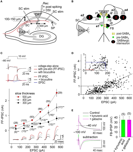
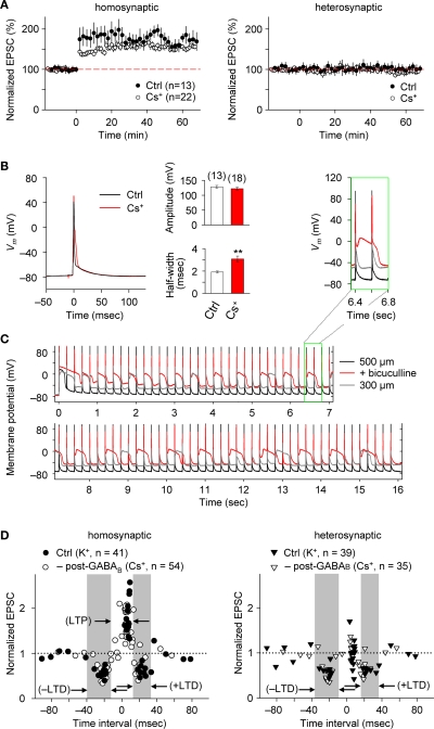
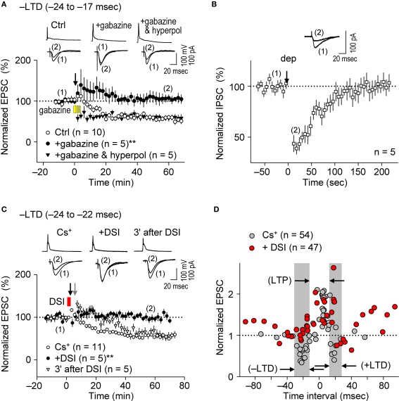
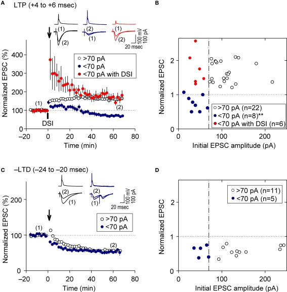
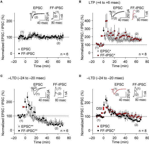
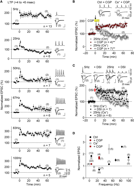
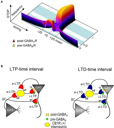
GABAergic interneuronal network activities in the hippocampus control a variety of neural functions, including learning and memory, by regulating θ and γ oscillations. How these GABAergic activities at pre- and postsynaptic sites of hippocampal CA1 pyramidal cells differentially contribute to synaptic function and plasticity during their repetitive pre- and postsynaptic spiking at θ and γ oscillations is largely unknown. We show here that activities mediated by postsynaptic GABA(A)Rs and presynaptic GABA(B)Rs determine, respectively, the spike timing- and frequency-dependence of activity-induced synaptic modifications at Schaffer collateral-CA1 excitatory synapses. We demonstrate that both feedforward and feedback GABA(A)R-mediated inhibition in the postsynaptic cell controls the spike timing-dependent long-term depression of excitatory inputs ("e-LTD") at the θ frequency. We also show that feedback postsynaptic inhibition specifically causes e-LTD of inputs that induce small postsynaptic currents (<70 pA) with LTP-timing, thus enforcing the requirement of cooperativity for induction of long-term potentiation at excitatory inputs ("e-LTP"). Furthermore, under spike-timing protocols that induce e-LTP and e-LTD at excitatory synapses, we observed parallel induction of LTP and LTD at inhibitory inputs ("i-LTP" and "i-LTD") to the same postsynaptic cells. Finally, we show that presynaptic GABA(B)R-mediated inhibition plays a major role in the induction of frequency-dependent e-LTD at α and β frequencies. These observations demonstrate the critical influence of GABAergic interneuronal network activities in regulating the spike timing- and frequency-dependences of long-term synaptic modifications in the hippocampus.

Keywords: CA1; GABA; STDP; frequency; hippocampal oscillation; spike timing.


Figures

Figure 1
Figure 1
Repetitive, single pre- and postsynaptic spiking recruits both FF and FB postsynaptic GABAAR-mediated inhibition in hippocampal CA1 pyramidal cells. (A) Experimental paradigm depicting the positions of stimulating and recording electrodes. Whole-cell recordings were made in the CA1 cell body and the test stimuli were applied at the stratum radiatum at least 500 μm distant from the recording electrode. (B) Schematic diagram of neuronal networks involved in the induction of STDP in CA1 pyramidal cells. Locations of excitatory (e) and inhibitory (i) inputs and GABA receptors are indicated. (C) Procedure for concurrent measurement of EPSCs and FF-IPSCs (top) and summary of their amplitudes (bottom). FF-IPSC is detected (top middle) with the use of voltage-steps (top upper) and presynaptic stimulation (pre-stim). The magnitude of FF-IPSC (top lower, black) is calculated by subtracting the voltage-step alone (top middle, black) from the pre-stimulation value (top middle, blue). The voltage-step at +10 mV was applied for 40 ms to prevent the induction of DSI (less than 1%; Lenz and Alger, 1999). The FF-IPSC was confirmed by GABAA blockade with bicuculline (20 μM, top middle and lower, red). The ratio of FF-IPSCs to EPSCs increased as the slice thickness increased. Data represent the mean EPSC/FF-IPSC (±sem). Traces: FF-IPSC in 500-μm (red), 400-μm (gray) and 300-μm (black) thick slices. The (number) indicates the total number of trials. Significant differences from corresponding data in 500-μm thick slices are indicated (“*” p < 0.05 and “**” p < 0.01; Mann–Whitney U test). (D) The amplitude of FF-IPSC is proportional to the EPSC amplitude. When the EPSC level is <70 pA, there is no FF-IPSC (see red line in inset). The EPSC amplitude normally used in this study is indicated (blue line in inset). (E) Prominent FB postsynaptic GABAAR-mediated inhibition is recruited by repetitive 5-Hz postsynaptic spiking in CA1 pyramidal cells. Postsynaptic spiking was induced by depolarizing current injections (6 nA, 2 ms, 20 pulses at 5 Hz) while postsynaptic CA1 pyramidal cells were held at −100 mV in a current-clamp configuration using a Cs+-based internal recording solution. The top left panel shows the postsynaptic membrane potentials monitored when FB postsynaptic GABAAR-mediated inhibition was blocked by either kynurenic acid (5 mM) or gabazine (10 μM). The control, in which GABAAR was not blocked, showed a feedback inhibition-inclusive membrane potential. Subtraction analysis (bottom left) revealed a membrane potential resulting from FB postsynaptic GABAAR-mediated inhibition. Average FB-IPSP (right).
Figure 2
Figure 2
e-STDP is induced at the θ frequency independently of postsynaptic GABABR function at SC-CA1 synapses. (A) Time course of changes in EPSC amplitudes induced by the LTP-timing protocol in homosynaptic, activated inputs (left) and heterosynaptic, non-activated inputs (right). Input-specific e-LTP was observed in K+-based (control, Ctrl) and Cs+-based internal recording solution. Data represent the normalized mean EPSC (±sem). (B) Action potentials recorded at the soma with the LTP time intervals in the presence or absence of K+ channel activities. Membrane potential (Vm) changes in either K+-based (black) or Cs+-based (red) internal solution (left). The average spike amplitude (right upper) and the half maximum width (right lower) were not as significantly different between the two experimental conditions as reported previously (Wittenberg and Wang, 2006). This may be due to extensive postsynaptic GABAAR-mediated inhibition in our preparation. Significant difference from the control is indicated (“**” p < 0.01; Mann–Whitney U test). (C) Membrane potentials (Vm) controlled by GABAAR-mediated inhibition. Stable Vm was observed in 500-μm (black), but not in 300-μm (gray) thick hippocampal slices in which elimination of postsynaptic GABAAR-mediated inhibition by bicuculline (20 μM) treatment caused depolarization (red). Inset: twice magnified view. (D) Summary of normalized EPSC changes in homosynaptic (left) and heterosynaptic (right) inputs. A similar timing dependence of e-STDP was observed in both K+-based (Ctrl) and Cs+-based (–post-GABAB; without postsynaptic GABABR-mediated inhibition) internal solution. e-LTD at both negative and positive time intervals propagated to heterosynaptic, non-activated synapses (right). Gray areas indicate e-LTD time intervals in the K+-based internal solution. Data recorded in the Cs+-based internal solution is adapted from (Nishiyama et al., 2000).
Figure 3
Figure 3
Postsynaptic GABAAR-mediated inhibition controls the spike-timing dependence of e-LTD at the θ frequency. (A) Bath application of gabazine (5 μM, yellow bars) abolished e-LTD (time intervals: −24 to −17 ms), whereas hyperpolarization (hyperpol) of postsynaptic cells restored e-LTD. (B) Brief postsynaptic depolarization (arrow; dep, 3 s) induced DSI at SC inputs. (C) DSI (red bar) induction immediately preceding the spike-timing protocol (black arrow) abolished e-LTD (time intervals: −24 to −22 ms), whereas DSI applied 3 min before (gray arrow) had no effect on the e-LTD. Data represent normalized mean EPSCs/IPSCs (±sem). Sample traces of membrane potentials during the spike-timing protocol (top, in A,C) and of IPSCs (in B)/EPSCs (bottom, in A,C) before (1) and after (2) the induction protocols. Significant differences from corresponding controls are indicated (“**” p < 0.01; Mann–Whitney U test). (D) Summary of time windows for e-STDP in the presence (filled red circle) and absence (filled gray circle) of DSI. Gray areas show LTD time intervals in the absence of DSI.
Figure 4
Figure 4
Feedback postsynaptic GABAAR-mediated inhibition causes e-LTD that enforces the requirement of cooperative e-LTP. (A,B) e-LTD is induced by the LTP-timing protocol when the initial EPSC amplitudes are <70 pA. DSI converts this e-LTD to e-LTP to the same extent as that induced normally at EPSC inputs of amplitude > 70 pA. (C,D) e-LTD is induced to a similar extent independent of the initial EPSC amplitudes. Significant difference from corresponding controls is indicated (“**” p < 0.01; Mann–Whitney U test).
Figure 5
Figure 5
Parallel induction of e-LTP/i-LTP and e-LTD/i-LTD occurs under prominent postsynaptic GABAAR-mediated inhibition. (A) Normalized EPSC/FF-IPSC amplitudes (%) without the spike-timing protocol. No changes in either EPSCs or FF-IPSCs were observed during the time course of the experiments. (B,C) EPSCs and FF-IPSCs were both potentiated at the LTP time intervals (B), and depressed at the –LTD time intervals (C). (D) DSI abolished LTD of EPSC, but not of FF-IPSC at the –LTD time intervals. Data represent normalized mean EPSCs/FF-IPSCs (±sem). Sample traces of EPSCs/FF-IPSCs (in B–D) before (1) and after (2) the induction protocols. Significant differences from corresponding controls are indicated (“*” p < 0.05 and “**” p < 0.01; Mann–Whitney U test).
Figure 6
Figure 6
Presynaptic GABABR-mediated inhibition causes frequency-dependent e-LTD at α/β frequencies. (A,D) Summary of frequency-dependent changes in synaptic efficacy induced by the spike-timing protocol for LTP-timing (+4 to +6 ms). Frequency-dependent effects (between 5 Hz and 100 Hz) in either the K+- or Cs+-based internal recording solution. Frequency-dependent e-LTD is induced at the same magnitude at 25 Hz and e-LTP is robust at the γ frequency in both recording solutions. (B) Bath application of CGP 35348 (1 mM, yellow bars) abolished e-LTD at 25 Hz, and instead induced e-LTP in both K+- and the Cs+-based internal recording solutions. (C) DSI failed to restore e-LTP induction at 25 Hz and had no significant effect on e-LTP at 5 Hz. Data represent normalized mean EPSCs (±sem). Membrane potentials during the spike-timing protocol (upper traces) and EPSCs (lower traces) before (1) and after (2) the induction protocols (in A–C). Scales: 100 pA (or 100 mV), 20 ms. Significant differences from corresponding controls are indicated (“**” p < 0.01; Mann–Whitney U test).
Figure 7
Figure 7
Model for STDP controlled by GABA functions in the CA1 network. (A) Both spike timing- and frequency-dependence of e-STDP and differential GABA function that gates e-LTD. (B) Parallel induction of e-LTP/i-LTP and e-LTD/i-LTD. Upon application of the LTP- (left) or LTD-timing (right) protocol at 5 Hz, all excitatory synapses undergo, respectively, either e-LTP or e-LTD. e-LTP or e-LTD in the CB1R-expressing interneuron (both at inputs e2 and e3) propagates passively to input i-1, respectively, as either i-LTP or i-LTD.

Similar articles

Cited by

References

    1. Bartos M., Vida I., Jonas P. (2007). Synaptic mechanisms of synchronized gamma oscillations in inhibitory interneuron networks. Nat. Rev. Neurosci. 8, 45–5610.1038/nrn2044 - DOI - PubMed
    1. Bi G.-q., Poo M.-m. (1998). Synaptic modifications in cultured hippocampal neurons: dependence on spike timing, synaptic strength, and postsynaptic cell type. J. Neurosci. 18, 10464–1047210.1016/S0031-9384(02)00933-2 - DOI - PMC - PubMed
    1. Bliss T. V., Collingridge G. L. (1993). A synaptic model of memory: long-term potentiation in the hippocampus. Nature 361, 31–3910.1038/361031a0 - DOI - PubMed
    1. Bracci E., Vreugdenhil M., Hack S. P., Jefferys J. G. R. (2001). Dynamic modulation of excitation and inhibition during stimulation at gamma and beta frequencies in the CA1 hippocampal region. J. Neurophysiol. 85, 2412–242210.1016/S0306-4522(02)00212-9 - DOI - PubMed
    1. Buzsáki G. (2002). Theta oscillations in the hippocampus. Neuron 33, 325–34010.1002/hipo.20113 - DOI - PubMed