. 2011 Mar 15;6(3):e17733.
doi: 10.1371/journal.pone.0017733.

Antitumor activity of Noscapine in combination with Doxorubicin in triple negative breast cancer

Affiliations

Antitumor activity of Noscapine in combination with Doxorubicin in triple negative breast cancer

Mahavir B Chougule et al. PLoS One. .

Abstract

Background: The aim of this study was to investigate the anticancer activity and mechanism of action of Noscapine alone and in combination with Doxorubicin against triple negative breast cancer (TNBC).

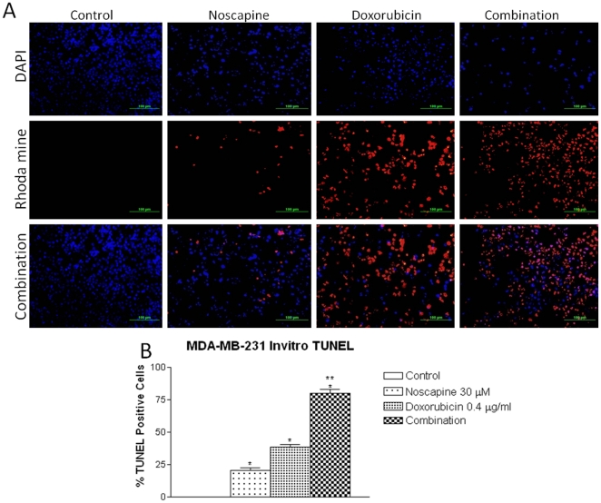
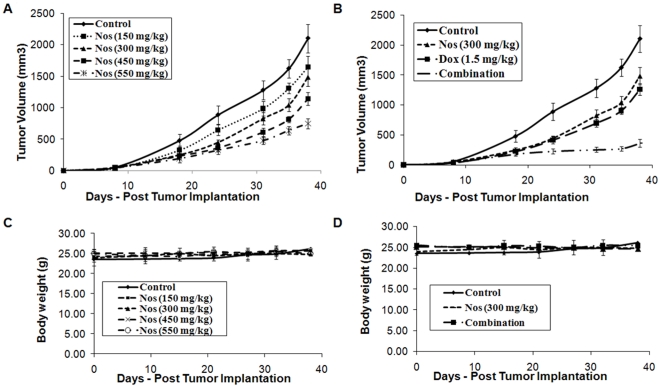
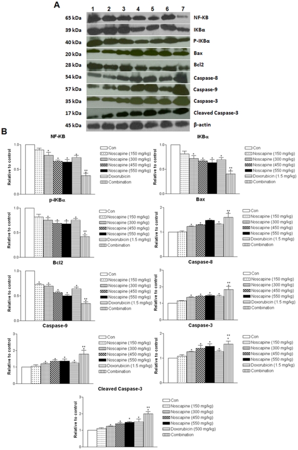
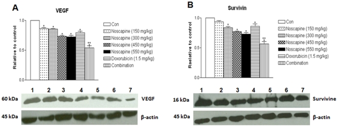
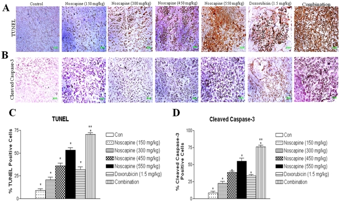
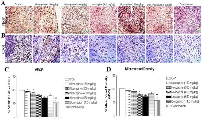
Methods: TNBC cells were pretreated with Noscapine or Doxorubicin or combination and combination index values were calculated using isobolographic method. Apoptosis was assessed by TUNEL staining. Female athymic Nu/nu mice were xenografted with MDA-MB-231 cells and the efficacy of Noscapine, Doxorubicin and combination was determined. Protein expression, immunohistochemical staining were evaluated in harvested tumor tissues.

Results: Noscapine inhibited growth of MDA-MB-231 and MDA-MB-468 cells with the IC(50) values of 36.16±3.76 and 42.7±4.3 µM respectively. The CI values (<0.59) were suggestive of strong synergistic interaction between Noscapine and Doxorubicin and combination treatment showed significant increase in apoptotic cells. Noscapine showed dose dependent reduction in the tumor volumes at a dose of 150-550 mg/kg/day compared to controls. Noscapine (300 mg/kg), Doxorubicin (1.5 mg/kg) and combination treatment reduced tumor volume by 39.4±5.8, 34.2±5.7 and 82.9±4.5 percent respectively and showed decreased expression of NF-KB pathway proteins, VEGF, cell survival, and increased expression of apoptotic and growth inhibitory proteins compared to single-agent treatment and control groups.

Conclusions: Noscapine potentiated the anticancer activity of Doxorubicin in a synergistic manner against TNBC tumors via inactivation of NF-KB and anti-angiogenic pathways while stimulating apoptosis. These findings suggest potential benefit for use of oral Noscapine and Doxorubicin combination therapy for treatment of more aggressive TNBC.


Conflict of interest statement

Competing Interests: The authors have declared that no competing interests exist.

Figures

Figure 1
Figure 1. Fluorescence Micrographs of cells stained with rhodamine and DAPI after 72 h (A) with Doxorubicin 0.4 µg/ml , Noscapine 30 µM, and, Noscapine and Doxorubicin combination in MDA-MB-231 cells and (B) Quantitation of apoptotic MDA-MB-231 cells from TUNEL assay.
DNA fragmentation indicated by positive staining (red) and nuclear condensation indicated by DAPI nuclear staining (blue). Micron bar = 100 µm. Cells were quantitated by counting 100 cells from 6 random microscopic fields. Data are expressed as mean+SD (N = 6). One-way ANOVA followed by post Tukey test was used for statistical analysis to compare control and treated groups. * P<0.01; all treatments significantly different from control and ** P<0.01; significantly different from Noscapine and Doxorubicin single treatments.
Figure 2
Figure 2. Progression profile of tumor growth kinetics of in-vivo antitumor effect of different doses of Noscapine alone (A) and in combination with Doxorubicin (B) on human MDA-MB-231 tumor xenograft model (tumor volumes, mm3 ± SEM), and measurement of body weight following Noscapine alone (C) and combination with Doxorubicin (D).
Female nude mice with xenograft MDA-MB-231 tumor tumors received various treatments for 38 days starting on day 7 post tumor implantation. The mice were treated with Noscapine (150–550 mg/kg/day), Doxorubicin 1.5 mg/kg i.v. bolus, q3d×7 schedule, and Noscapine 300 mg/kg/day+Doxorubicin 1.5 mg/kg i.v. bolus, q3d×7 schedule. Control group received vehicle only. Statistical significance of the difference in tumor volume of treatment groups compared with control. P<0.01 (*, significantly different from untreated controls; **, significantly different from Noscapine and Doxorubicin single treatments). Data presented are means and SE (n = 8). This experiment was repeated twice.
Figure 3
Figure 3. Western blotting of tumor tissue lysates to determine expressions apoptosis-related proteins (A) expression of NF-kβ, IKBα, P-IKBα, Bax, Bcl2, caspase 3, cleaved caspase 3, activated caspase 8 and activated caspase 9 proteins in tumor lysates by western blotting and (B) quantitation of apoptotic protein expression.
Tumor tissue lysates harvested tumor tissues from control-untreated and treated groups were analyzed by western blotting for protein expressions. Lane 1 = control; Lane 2 = Noscapine 150 mg/kg/day; Lane 3 = Noscapine 300 mg/kg/day; Lane 4 = Noscapine 450 mg/kg/day; Lane 5 = Noscapine 550 mg/kg/day; Lane 6 = Doxorubicin 1.5 mg/kg i.v. bolus, q3d×7 schedule; Lane 7 = Combination (Noscapine 300 mg/kg/day+Doxorubicin1.5 mg/kg i.v. bolus, q3d×7 schedule). Similar results were observed in triplicate experiments. Protein expression levels (relative to β-actin) were determined. Mean ± SE for three replicate determinations. One-way ANOVA followed by post Tukey test was used for statistical analysis. P<0.01 (*, significantly different from untreated controls; **, significantly different from Noscapine and Doxorubicin single treatments).
Figure 4
Figure 4. Western blotting of tumor tissue lysates to determine expressions angiogenesis-related proteins expression of (A) VEGF and (B) survivin proteins in tumors and quantitation of protein expression.
Whole-cell lysates from control-untreated and treated tumors were analyzed by western blotting for protein expressions. Lane 1 = control; Lane 2 = Noscapine 150 mg/kg/day ; Lane 3 = Noscapine 300 mg/kg/day ; Lane 4 = Noscapine 450 mg/kg/day; Lane 5 = Noscapine 550 mg/kg/day; Lane 6 = Doxorubicin 1.5 mg/kg i.v. bolus, q3d×7 schedule; Lane 7 = Combination (Noscapine 300 mg/kg/day+Doxorubicin ). Similar results were observed in replicate experiments. Protein expression levels (relative to β-actin) were determined. Mean ± SE for three replicate determinations. One-way ANOVA followed by post Tukey test was used for statistical analysis. P<0.01 (*, significantly different from untreated controls; **, significantly different from Noscapine and Doxorubicin single treatments).
Figure 5
Figure 5. Immunohistochemical staining of xenograft MDA-MB-231 breast tumor tissues for induction of apoptosis using TUNEL assay (A); for expression of cleaved caspase 3 (B); quantitation of apoptotic cells from TUNEL staining (C); and quantitation of caspase 3 positive cells apoptotic cells (D).
Tumor sections were stained using the DeadEnd colorimetric kit and cleaved caspase-3 (Asp 175) IHC kit for TUNEL assay and cleaved caspase 3 expression as described in materials and methods respectively. The apoptotic tumor cells are stained brown. Percentages of TUNEL-positive and cleaved caspase 3-positive cells were quantitated by counting 100 cells from 6 random microscopic fields. Data are expressed as mean+SD (N = 6). One-way ANOVA followed by post Tukey test was used for statistical analysis to compare control and treated groups. P<0.01 (*, significantly different from untreated controls; **, significantly different from Noscapine and Doxorubicin single treatments). Original magnification ×40 (Micron bar = 100 µm).
Figure 6
Figure 6. Immunohistochemical staining of MDA-MB-231 tumor tissues for (A) VEGF expression.
Tumor sections were stained using the ABC staining kit as described in Materials and Methods. Cells showing positive VEGF expression are stained brown. Original magnification ×40. (Micron bar = 100 µm). Immunohistochemical staining of MDA-MB-231 tumor tissues for (B) CD31 expression. Tumor angiogenesis was assessed by immunohistochemical staining with anti-CD31 antibody (brown) on paraffin-embedded sections. Original magnification ×40. (Micron bar = 100 µm). (C) Quantitation of apoptotic cells from VEGF staining. (D) Assessment of microvessel density. Microvessel density (MVD) was calculated by selecting three most vascularised areas of the tumour (‘hot spots’) and mean values obtained by counting vessels. A single microvessel was defined as a discrete cluster of cells positive for CD31 staining, with no requirement for the presence of a lumen. Microvessel counts were performed at ×400 (×40 objective lens and ×10 ocular lens; 0.74 mm2 per field). The MVD was significantly different between the control group and treated groups in sequential analysis; **, P<0.01;* P<0.05 relative to control.

References

    1. Kassam F, Enright K, Dent R, Dranitsaris G, Myers J, et al. Survival outcomes for patients with metastatic triple-negative breast cancer: implications for clinical practice and trial design. Clin Breast Cancer. 2009;9:29–33. - PubMed
    1. Irvin WJ, Jr, Carey LA. What is triple-negative breast cancer? Eur J Cancer. 2008;44:2799–2805. - PubMed
    1. Stockmans G, Deraedt K, Wildiers H, Moerman P, Paridaens R. Triple-negative breast cancer. Curr Opin Oncol. 2008;20:614–620. - PubMed
    1. Moulder S, Hortobagyi GN. Advances in the treatment of breast cancer. Clin Pharmacol Ther. 2008;83:26–36. - PubMed
    1. Buzdar AU. Advances in endocrine treatments for postmenopausal women with metastatic and early breast cancer. Oncologist. 2003;8:335–341. - PubMed

Publication types

MeSH terms