. 2011 Mar 14;6(3):e17739.
doi: 10.1371/journal.pone.0017739.

IL-21 and IL-6 are critical for different aspects of B cell immunity and redundantly induce optimal follicular helper CD4 T cell (Tfh) differentiation

Affiliations

IL-21 and IL-6 are critical for different aspects of B cell immunity and redundantly induce optimal follicular helper CD4 T cell (Tfh) differentiation

Danelle Eto et al. PLoS One. .

Abstract

Cytokines are important modulators of lymphocytes, and both interleukin-21 (IL-21) and IL-6 have proposed roles in T follicular helper (Tfh) differentiation, and directly act on B cells. Here we investigated the absence of IL-6 alone, IL-21 alone, or the combined lack of IL-6 and IL-21 on Tfh differentiation and the development of B cell immunity in vivo. C57BL/6 or IL-21(-/-) mice were treated with a neutralizing monoclonal antibody against IL-6 throughout the course of an acute viral infection (lymphocytic choriomeningitis virus, LCMV). The combined absence of IL-6 and IL-21 resulted in reduced Tfh differentiation and reduced Bcl6 protein expression. In addition, we observed that these cytokines had a large impact on antigen-specific B cell responses. IL-6 and IL-21 collaborate in the acute T-dependent antiviral antibody response (90% loss of circulating antiviral IgG in the absence of both cytokines). In contrast, we observed reduced germinal center formation only in the absence of IL-21. Absence of IL-6 had no impact on germinal centers, and combined absence of both IL-21 and IL-6 revealed no synergistic effect on germinal center B cell development. Studying CD4 T cells in vitro, we found that high IL-21 production was not associated with high Bcl6 or CXCR5 expression. TCR stimulation of purified naïve CD4 T cells in the presence of IL-6 also did not result in Tfh differentiation, as determined by Bcl6 or CXCR5 protein expression. Cumulatively, our data indicates that optimal Tfh formation requires IL-21 and IL-6, and that cytokines alone are insufficient to drive Tfh differentiation.


Conflict of interest statement

Competing Interests: The authors have declared that no competing interests exist.

Figures

Figure 1
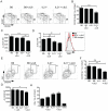
Figure 1. IL-21 can be produced by Th1, Th2 or Th17 cell in vitro.
(A–D) Magnetic bead purified total CD4 T cells were stimulated with plate-bound αCD3 plus αCD28 without cytokine blocking mAbs (unbiased, U), or in the presence of αIFNγ + αIL-4 (Th0), IL-12 + αIL-4 (Th1), IL-4 + αIFNγ (Th2), or IL-6/TGFβ + αIFNγ & αIL-4 (Th17). Day 7 CD4+B220 gated in vitro cultured cells were stained by ICS after 4 h stimulation in PMA/ionomycin. Representative dot plots of IL-21 and the respective cytokine associated with each subset, (A) IFNγ (Th1), (B) IL-4 (Th2), or (C) IL-17 (Th17). (D) IL-21 was measured by ELISA from day 7 cells that had been washed, and then restimulated overnight in the presence of PMA/ionomycin. IL-21 was normalized to cell number per well. (E) Sorted naïve CD4 T cells (CD4+CD44NK1.1CD25) were cultured under unbiased (U), Th0 or Th1 conditions. ELISA analysis of IL-21 production from day 7 cultured CD4 T cells restimulated overnight with PMA/ionomycin (normalized to cell number per well). “—”  =  no stim. Data are representative of ≥2 independent experiments.
Figure 2
Figure 2. In vitro stimulation of purified naïve CD4 T cells with IL-6 or IL-21 drives high IL-21 expression.
(A–D) Naïve sorted CD4 T cells (CD4+CD44NK1.1CD25) were stimulated for 3 days on αCD3/αCD28 coated plates in the presence of αIFNγ, αIL-4, and αTGFβ (Th0) ± IL-6 or IL-21. Reagents to block IL-21 (5 µg/ml) or IL-6 (10 µg/ml) signaling were also added at this time where indicated. IL-21R-Fc intracellular staining was used to identify IL-21 producing cells after 4 h of PMA/ionomycin stimulation. (A) Flow cytometry analysis of IL-21 and IFNγ production in day 7 Th0, Th0 + IL-6, and Th0 + IL-6 + αIL6 polarized CD4 T cell cultures. Cells were gated on B220CD44hi CD4 T cells. *** P<0.0005. (B–D) FACS analysis of IL-21 and IFNγ production in day 3 Th0 + IL-21± αIL-21 or IL-21R-Fc differentiated CD4 T cells. (B) Representative plots (B220CD44hi CD4 T cells). (C) Quantitation of IL-21 production or (D) IL-21 MFI. ** P<0.003, *** P<0.0003. Data are representative of ≥2 independent experiments for each condition with duplicate samples. MFI, mean fluorescence intensity.
Figure 3
Figure 3. High IL-21 production in CD4 T cell cultures does not instruct Tfh differentiation.
Naïve sorted CD4 T cells were stimulated for 72 h on αCD3 plus αCD28 coated plates in the presence of αIFNγ, αIL-4, and αTGFβ (Th0) ± IL-6 or IL-21. (A) CXCR5 mRNA expression normalized to a housekeeping gene, G6PDH, in naïve (CD4+CD44lo), day 3 in vitro polarized CD4 T cell cultures (Th0 or Th0 + IL-6), or OTII CD4 T cells from day 8 NP-OVA plus alum in vivo immunized mice (CD44hiCXCR5+GL7 Tfh or CD44hiCXCR5+GL7+ GC Tfh). (B) Quantitation of CXCR5 mean fluorescence intensity (MFI) in day 3 cultures (Th0 or Th0 + IL-6) or CD4 splenocytes (naïve, CD44lo; CXCR5+, CD44hi), and histogram. (C) CXCR5 analysis in 72 h Th0 (black) or Th0 + IL-21 (red) cultures. Th0 and Th0 + IL-21 cultures stained with isotype mAb (blue). (D–E) Timecourse of Bcl6 (D) and Blimp-1 (E) mRNA expression normalized to a housekeeping gene, G6PDH, in Th0 or Th0 + IL-6 CD4 T cell cultures. (F) FACS analysis of Bcl6 protein expression in germinal center B cells (GL7+B220+) versus non GC B cells (GL7B220+) in splenocytes 8 days following LCMV infection. (G) Bcl6 protein expression in day 3 in vitro differentiated CD4 cultures (Th0 or Th0 + IL-6) versus freshly isolated naïve CD4 splenocytes (CD44lo). Left, quantitation of Bcl6 MFI. Right, Bcl6 histogram. (H) Bcl6 analysis in 72 h Th0 (black) or Th0 + IL-21 (red) cultures. Naïve splenocytes are stained for comparison (CD44lo, gray). Data are representative of ≥2 independent experiments with duplicate samples.
Figure 4
Figure 4. Cooperation of IL-6 and IL-21 for plasma cell generation and antibody responses.
C57BL/6 (B6) or IL-21−/− mice treated with αIL-6 or isotype mAb throughout the acute LCMV infection. (A) Titers of anti-LCMV IgG in the serum of B6 or IL-21−/− mice ± αIL-6 mAb 8 days following LCMV infection: B6 vs. B6 + αIL-6 (**, P = 0.0016), B6 vs. IL-21−/− (*, P = 0.025), B6 vs. IL-21−/− + αIL-6 (**, P = 0.0016), and IL-21−/− vs. IL-21−/− + αIL-6 (**, P = 0.0012). (B) Quantitation of plasma cell development by FACS: B6 vs. IL-21−/− (***, P = 0.0008), B6 vs. IL-21−/− + αIL-6 (***, P<0.0001), and IL-21−/− ± anti-IL-6 (*, P = 0.0143). (C) Plasma cell (CD138+IgD) development 8 days post-infection, gated on CD19+ splenocytes. Data are representative of 3 independent experiments; n = 5–6 mice per group. ns, not significant.
Figure 5
Figure 5. IL-21, but not IL-6, is needed for optimal germinal center B cell development.
(A) Germinal center B cells (GL7+Fas+ gated, total B cell gate shown) in C57BL/6 (B6) or IL-21−/− mice treated with anti-IL-6 (αIL6) or isotype mAb. Splenocytes were analyzed 8 days following acute LCMV infection. (B) Frequency of germinal center B cells of total B cells. *, P<0.02. (C) Quantitation of the PNA mean fluorescence intensity (MFI) in germinal center B cells. ***, P = 0.0003. Data are representative of 3 independent experiments; n = 5–6 mice per group. MFI, mean fluorescence intensity; ns, not significant.
Figure 6
Figure 6. IL-6 or IL-21 is required for optimal Tfh differentiation.
(A–G) Splenocytes were analyzed 8 days following LCMV infection in C57BL/6 (B6) or IL-21−/− mice ± αIL6. (A) Follicular helper T (Tfh) cells (CXCR5+SLAMlo, boxed) gated on activated (CD44hi) CD4 T cells. (B) Percentage of Tfh differentiation (from gated Tfh population). **, P = 0.0016. (C) Quantitation of ΔCXCR5 mean fluorescence intensity (MFI) gated on Tfh (normalized against CD44lo CD4 T cells). **, P = 0.003. ***, P<0.0001. (D) Left, quantitation of Bcl6 protein ΔMFI in Tfh (normalized against CD44lo CD4 T cells). ***, P = 0.0009. Right, histogram overlay shows Bcl6 expression in a representative B6 vs. IL-21−/− + αIL-6 Tfh population (B6, black; IL-21−/− + αIL-6, red; B6 stained with isotype mAb, gray). (E) FACS analysis of germinal center CD4 T (GC Tfh) cells (CXCR5+GL7+, boxed). Gated CD44hi CD4 T cells shown. (F) GC Tfh quantitation. **, P = 0.003. (G) ΔCXCR5 MFI, gated on GC Tfh and normalized against CD44lo CD4 T cells. *, P = 0.0405. Flow cytometric analysis of intracellular cytokine staining for IFNγ production by CD4 T cells after 4 h stimulation with PMA and ionomycin in the presence of brefeldin A. (H) Frequency of IFNγ+CD44hi CD4 T cells. Data are representative of ≥2 independent experiments; n = 5–6 mice per group. MFI, mean fluorescence intensity; ns, not significant.

Similar articles

Cited by

References

    1. Crotty S, Ahmed R. Immunological memory in humans. Semin Immunol. 2004;16:197–203. - PubMed
    1. MacLennan IC. Germinal centers. Annu Rev Immunol. 1994;12:117–139. - PubMed
    1. Allen CDC, Okada T, Cyster JG. Germinal-center organization and cellular dynamics. Immunity. 2007;27:190–202. - PMC - PubMed
    1. Johnston RJ, Poholek AC, DiToro D, Yusuf I, Eto D, et al. Bcl6 and Blimp-1 are reciprocal and antagonistic regulators of T follicular helper cell differentiation. Science. 2009;325:1006–1010. - PMC - PubMed
    1. Nurieva RI, Chung Y, Martinez GJ, Yang XO, Tanaka S, et al. Bcl6 mediates the development of T follicular helper cells. Science. 2009;325:1001–1005. - PMC - PubMed

Publication types

MeSH terms