. 2011 Mar 14;6(3):e17732.
doi: 10.1371/journal.pone.0017732.

Conjugation to the cell-penetrating peptide TAT potentiates the photodynamic effect of carboxytetramethylrhodamine

Affiliations

Conjugation to the cell-penetrating peptide TAT potentiates the photodynamic effect of carboxytetramethylrhodamine

Divyamani Srinivasan et al. PLoS One. .

Abstract

Background: Cell-penetrating peptides (CPPs) can transport macromolecular cargos into live cells. However, the cellular delivery efficiency of these reagents is often suboptimal because CPP-cargo conjugates typically remain trapped inside endosomes. Interestingly, irradiation of fluorescently labeled CPPs with light increases the release of the peptide and its cargos into the cytosol. However, the mechanism of this phenomenon is not clear. Here we investigate the molecular basis of the photo-induced endosomolytic activity of the prototypical CPPs TAT labeled to the fluorophore 5(6)-carboxytetramethylrhodamine (TMR).

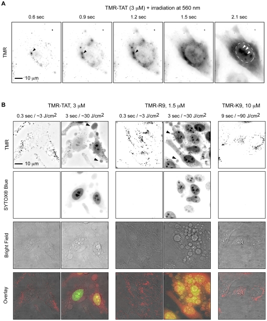
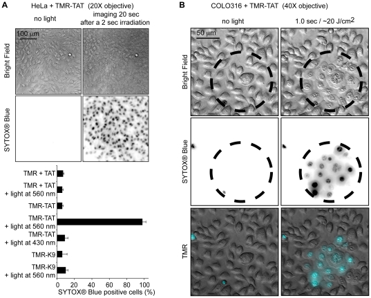
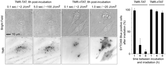
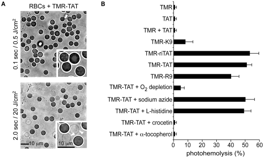
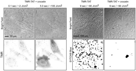
Methodology/principal findings: We report that TMR-TAT acts as a photosensitizer that can destroy membranes. TMR-TAT escapes from endosomes after exposure to moderate light doses. However, this is also accompanied by loss of plasma membrane integrity, membrane blebbing, and cell-death. In addition, the peptide causes the destruction of cells when applied extracellularly and also triggers the photohemolysis of red blood cells. These photolytic and photocytotoxic effects were inhibited by hydrophobic singlet oxygen quenchers but not by hydrophilic quenchers.

Conclusions/significance: Together, these results suggest that TAT can convert an innocuous fluorophore such as TMR into a potent photolytic agent. This effect involves the targeting of the fluorophore to cellular membranes and the production of singlet oxygen within the hydrophobic environment of the membranes. Our findings may be relevant for the design of reagents with photo-induced endosomolytic activity. The photocytotoxicity exhibited by TMR-TAT also suggests that CPP-chromophore conjugates could aid the development of novel Photodynamic Therapy agents.


Conflict of interest statement

Competing Interests: The authors have declared that no competing interests exist.

Figures

Figure 1
Figure 1. Photolytic effects of TMR-TAT after endocytosis in HeLa cells.
A) Light irradiation causes escape of TMR-TAT from endocytic organelles into the cytosol. Hela cells were incubated with TMR-TAT (3 µM), washed, and irradiated at 560±20 nm through a 100× objective on a wide-field microscope. Images were acquired in a time-lapse experiment with a light excitation of 300 ms and an interval of 2 seconds. The time displayed on the images represents the total light exposure time. The TMR fluorescence signal is represented as inverted monochrome (black = fluorescent signal, white = no signal). The TMR signal, initially in a punctate distribution, can be seen to diffuse away from individual endocytic organelles upon irradiation (black arrows). The perimeter of the nucleus is highlighted by a dashed line in the last image and the signal from TMR-TAT presumably accumulated at nucleoli is indicated with white arrows. B) Photosensitization of TMR-TAT or TMR-R9 endocytosed by cells causes plasma membrane damage and cell death. In contrast, cells containing TMR-K9 remain viable and the punctate distribution of TMR-K9 is not affected by the light irradiation under the conditions tested. The fluorescence signals of TMR and SYTOX® Blue are represented as inverted monochrome or pseudo-colored red and green in the overlay image, respectively.
Figure 2
Figure 2. The photolytic and photocytotoxic effects of TMR-TAT occur rapidly and in all irradiated cells.
A) Irradiation of TMR-TAT endocytosed by cells causes plasma membrane blebbing and permeabilization (SYTOX® Blue staining) within 20 seconds after 2 sec irradiation at 560 nm(the radiant exposure is approximately 40 J/cm2). Cell destruction is observed in more than 95% of the cells irradiated at 560 nm. In contrast, no photocytoxicity is observed when cells treated with TMR-K9 (10 µM) or TMR (3 µM) and TAT (3 µM) are irradiated under similar conditions. Cells incubated with TMR-TAT but irradiated at 430 nm are not destroyed (excitation wavelength of SYTOX® Blue but not of TMR, radiant exposure was also approximately 40 J/cm2). The histogram represents the average percentage of cells stained by SYTOX® Blue after peptide and light treatment (the number of cells examined is at least 3000) and the error bars represent the standard deviation (experiments were reproduced at least 3 times). B) The cytotoxic effect of TMR-TAT is limited to only irradiated areas. COLO 316 cells were incubated with TMR-TAT (3 µM) for 1 h. The cells were washed with fresh media and endocytosis of TMR-TAT was confirmed by fluorescence microscopy. The cells within the circled area were exposed to light at 560 nm. Morphological changes and SYTOX® Blue staining (inverted monochrome or pseudo-colored cyan) are only observable in the irradiated area.
Figure 3
Figure 3. Degradation of TMR-TAT in endocytic organelles abolishes photo-induced endosomal release and photocytotoxicity.
HeLa cells were incubated with TMR-TAT or TMR-riTAT (3 µM) for 1 h, washed, and incubated at 37°C for an additional 1, 4 or 8 hours. Irradiation of cells incubated with TMR-riTAT led to the cytosolic distribution of the TMR fluorescence signal, plasma membrane blebbing (as seen in the bright field image) and permeabilization (represented in histogram) under all tested conditions. In contrast, these photo-induced effects are dramatically reduced for TMR-TAT as the time between peptide incubation and irradiation is increased. The histogram represents the average percentage of cells stained by SYTOX® Blue after peptide and light treatment (the number of cells examined is at least 3000) and the error bars represent the standard deviation (experiments were reproduced at least 3 times).
Figure 4
Figure 4. Photosensitization of extracellular TMR-TAT causes plasma membrane blebbing, plasma membrane permeabilization, and cell shrinkage.
A) HeLa cells were incubated with TMR-TAT (3 µM) at 4°C to block endocytosis of the peptide. Images represent the bright field image and the fluorescence image of SYTOX® Blue (inverted monochrome). B) The effects of photosensitization of hematoporphyrin (HP) on cell morphology are similar to those obtained by photosensitization of TMR-TAT. HP was incubated with HeLa cells for 1 hour, washed with fresh L-15 media, and irradiated at 560 nm. Black arrows in A) and B) highlight the membrane blebbing observed during light exposure.
Figure 5
Figure 5. Photosensitization of TMR-TAT causes the lysis of red blood cells by the production of singlet oxygen in their membrane.
A) RBCs incubated with TMR-TAT (2 µM) have either a concave or crenated morphology initially. Irradiation of the sample at 560 nm causes formation of spherical cell ghosts (highlighted with white arrows in inserts) that lose their contrast in bright field images as the cells lyse. B) Percentage of lysed RBCs as a function of the compounds present in the media. TMR and TMR containing peptides were used at 2 µM. All experiments were performed by irradiating the samples at 560 nm for 400 msec (∼8 J/cm2). The data in the histogram represents the average of 4 experiments and the error bars correspond to the standard deviation. No lysis was observed for any of the samples in the absence of light.
Figure 6
Figure 6. Crocetin inhibits photosensitization of TMR-TAT.
TMR-TAT (3 µM) was incubated with crocetin (50 µM) and HeLa cells for 1 hour. Cells were washed with fresh L-15, placed on the microscope, and irradiated at 560 nm for the times indicated. The left panel represents cells imaged with the 100× and illustrates that endosomal release of TMR-TAT appears reduced by crocetin but not abolished. However, endosomal release is not accompanied by membrane blebbing or SYTOX® Blue staining (no signal detected, not shown) as seen when crocetin is omitted (right panel, cells are imaged with 20× objective).

References

    1. Zorko M, Langel U. Cell-penetrating peptides: mechanism and kinetics of cargo delivery. Adv Drug Deliv Rev. 2005;57:529–545. - PubMed
    1. Maiolo JR, 3rd, Ottinger EA, Ferrer M. Specific redistribution of cell-penetrating peptides from endosomes to the cytoplasm and nucleus upon laser illumination. J Am Chem Soc. 2004;126:15376–15377. - PubMed
    1. Matsushita M, Noguchi H, Lu YF, Tomizawa K, Michiue H, et al. Photo-acceleration of protein release from endosome in the protein transduction system. FEBS Lett. 2004;572:221–226. - PubMed
    1. Endoh T, Sisido M, Ohtsuki T. Spatial regulation of specific gene expression through photoactivation of RNAi. J Control Release. 2009;137:241–245. - PubMed
    1. Woods LK, Morgan RT, Quinn LA, Moore GE, Semple TU, et al. Comparison of four new cell lines from patients with adenocarcinoma of the ovary. Cancer Res. 1979;39:4449–4459. - PubMed

Publication types

MeSH terms