. 2010 Nov 1;6(4):266-281.
doi: 10.2174/157340210793611668.

Central Arterial Aging and Angiotensin II Signaling

Affiliations

Central Arterial Aging and Angiotensin II Signaling

Mingyi Wang et al. Curr Hypertens Rev. .

Abstract

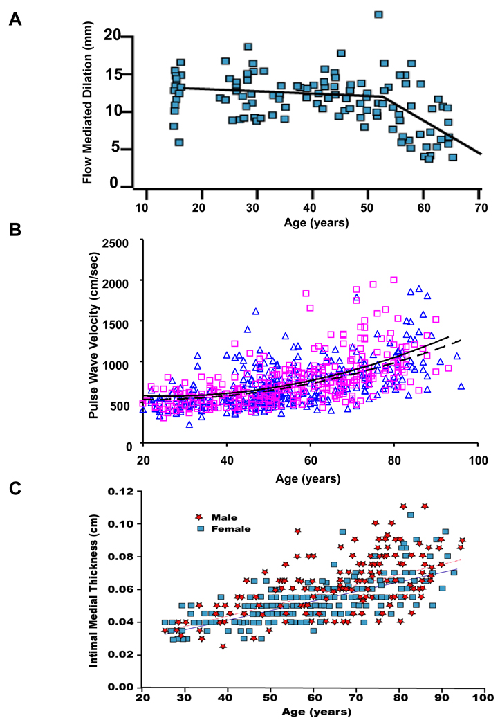
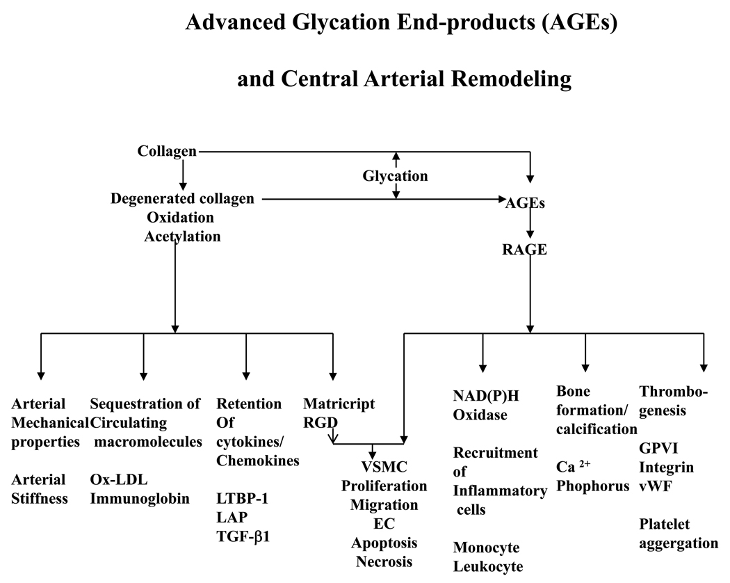
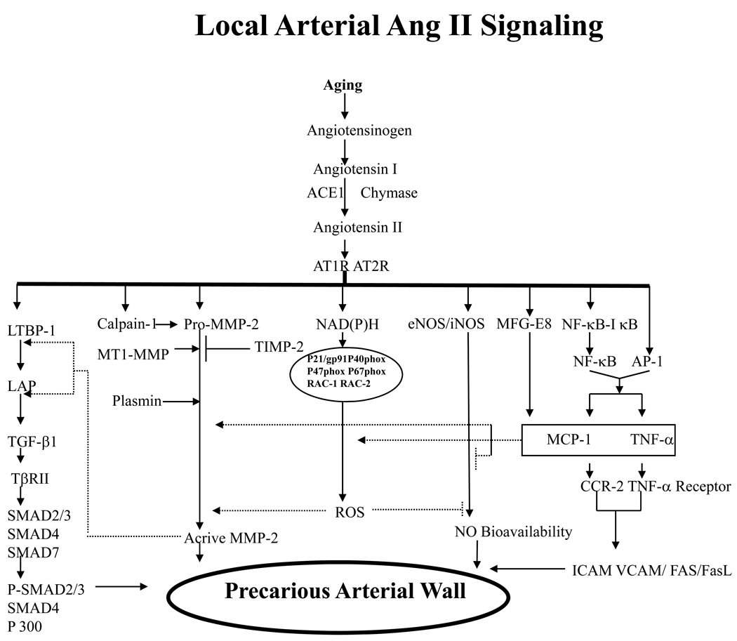
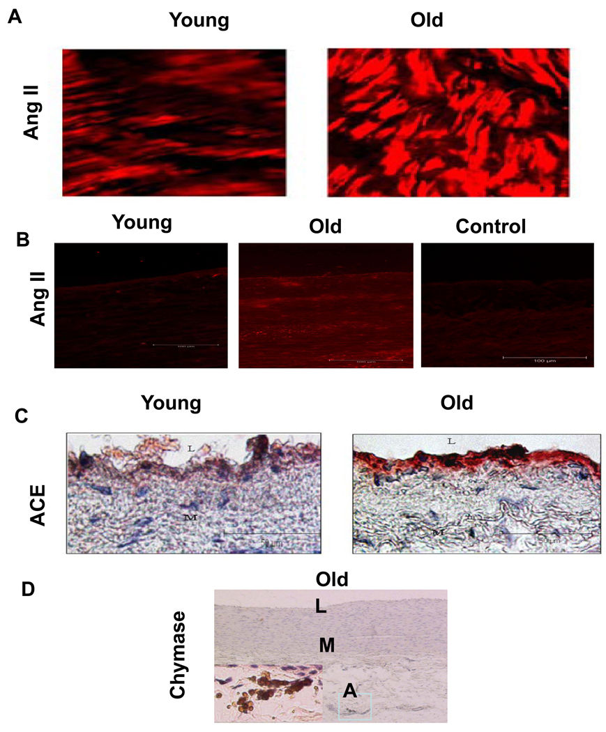
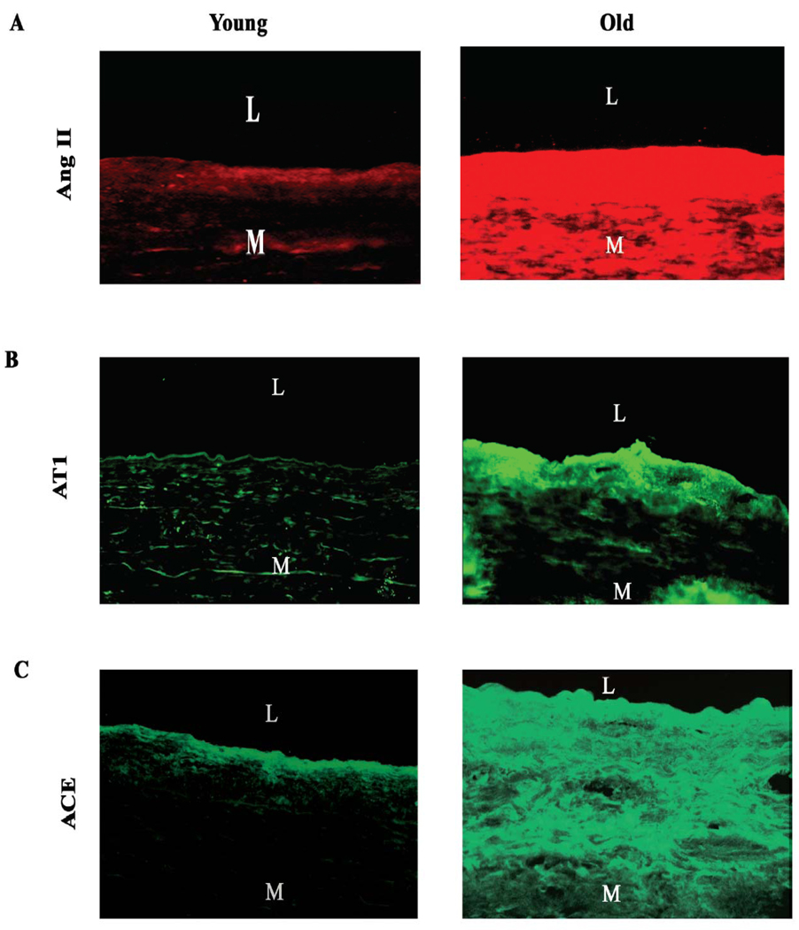
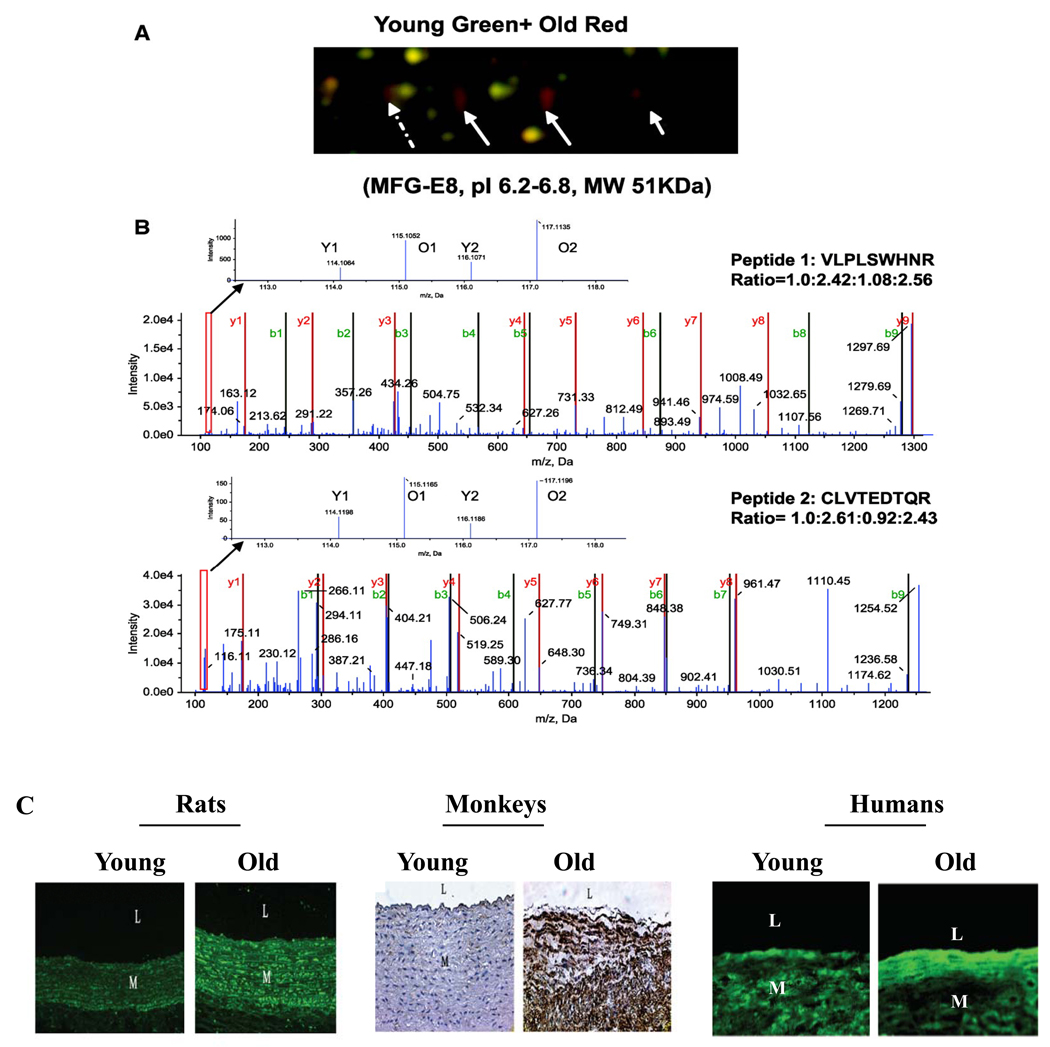
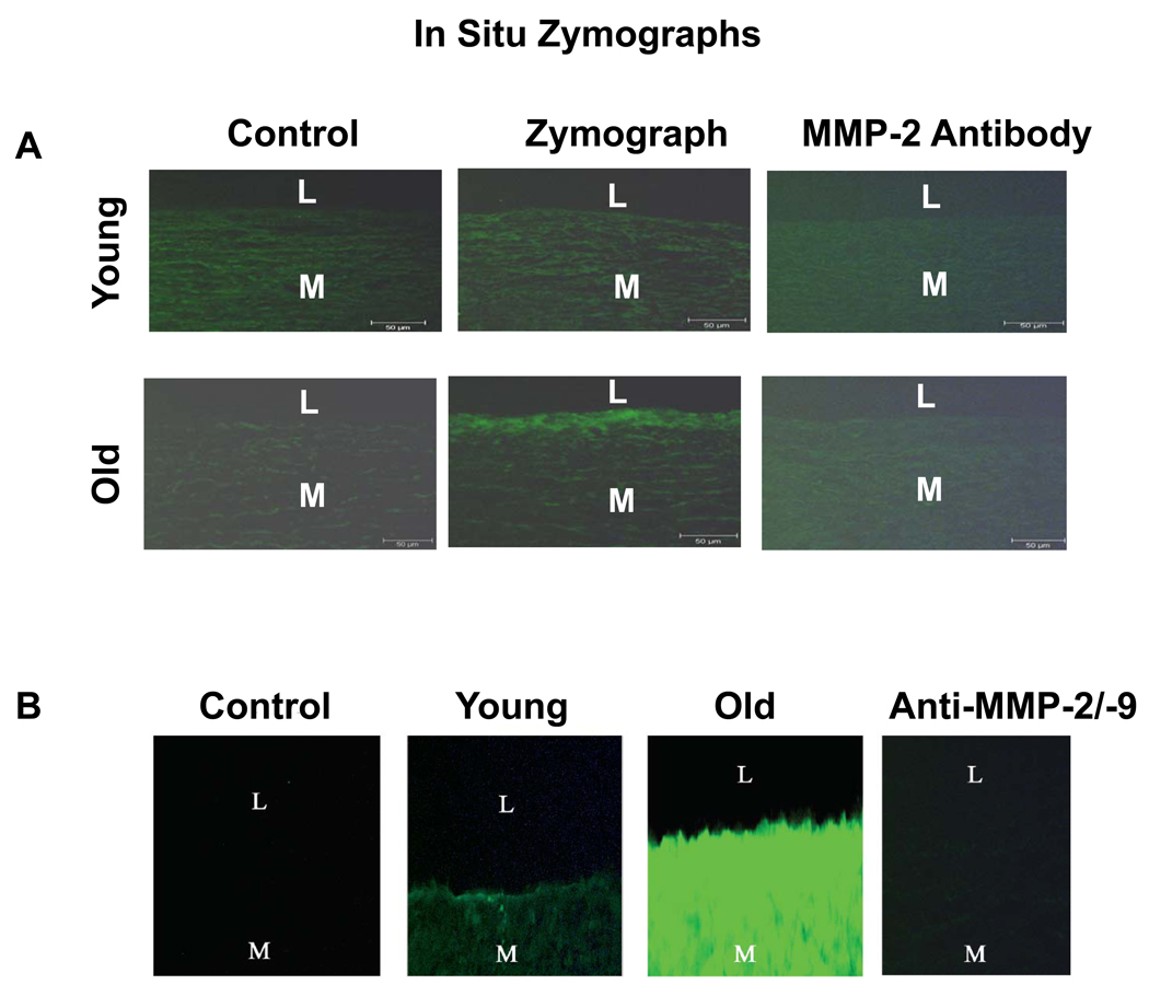
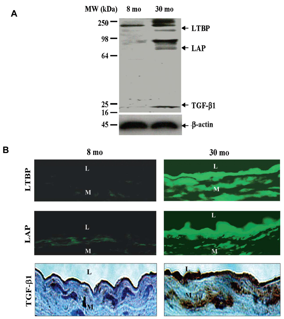
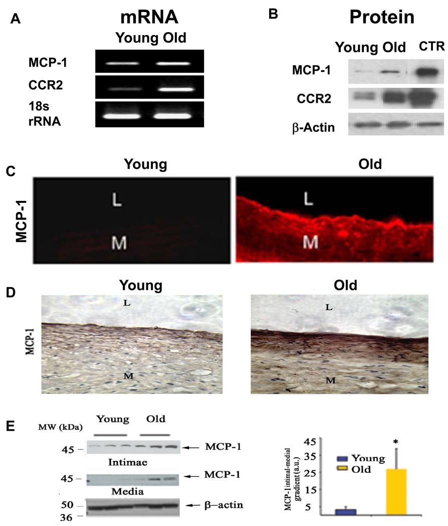
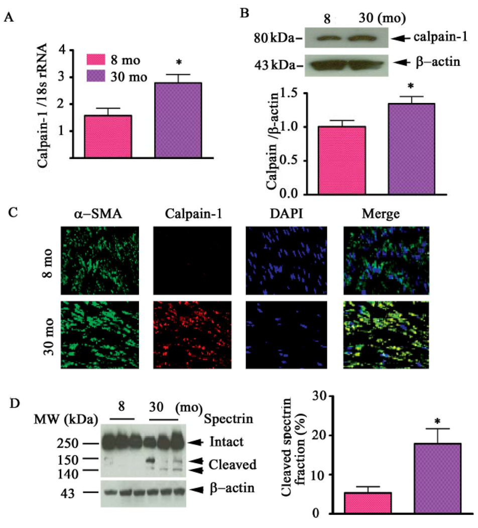
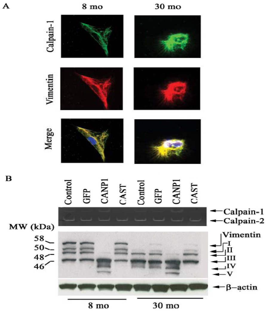
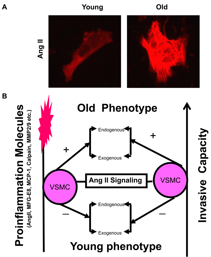
Arterial remodeling over time is a cornerstone of normal systemic aging. The age-associated arterial structural and functional changes in the intima, the media, and the adventitia are closely linked to angiotensin II (Ang II) signaling. A growing line of evidence indicates that essential elements of Ang II signaling, which encompasses milk fat globule epidermal growth factor-8, calpain-1, transforming growth factor-β1, matrix metalloproteinase-2/9, monocyte chemoattractant protein-1, nicotinamide adenine dinucleotide phosphate-oxidase, and reactive oxygen species, are upregulated within the central arterial wall in rats, nonhuman primates, and humans during aging. In vitro studies show that the elevation of Ang II signaling induces the accumulation of collagen and advanced glycated end-products, the degradation of elastin, and the increased cell cycle disorder, invasion, and hypertrophy of endothelial and vascular smooth muscle cells. Further, in vivo studies demonstrate that increased Ang II signaling accelerates arterial aging. Conversely, attenuating Ang II signaling via an inhibition of angiotensin conversing enzyme or a blockade of AT1 activation retards age-associated arterial remodeling. This review attempts to integrate complex facts of Ang II signaling within the aged central arterial wall and may shed light on new therapeutic targets for arterial aging.


Conflict of interest statement

Conflicts of Interest

None

Figures

Figure 1
Figure 1. Age-associated changes in arterial function in humans
A. Flow mediated induced dilation in the brachial artery of apparently healthy men. From Celermajer et al [15]. Age-associated increase in carotid-femoral pulse wave velocity (B), an index of central arterial stiffness, and intimal medial thickness (C) in healthy men (red) and women (blue). Best-fitting age regression curves are shown for men (solid lines) and women (dashed lines). From Vaitkevicius PV et al [16] and Nagai Y et al [18].
Figure 2
Figure 2
A schematic depiction of the effects of AGEs and their potential role in arterial remodeling. From Wang M et al [52].
Figure 3
Figure 3
Simplified schematic of the pleiotropic roles of increased Ang II signaling on arterial vulnerability. An age-associated increase in Ang II induces TGF-β expression, activates the nuclear factor κB (NF-κB) and MMP systems, promotes reactive oxygen species (ROS) production, and decreases NO bioavailability, contributing to arterial inflammation and fibrosis and resulting in arterial remodeling. ACE indicates angiotensin-converting enzyme; AT1R, Ang II type 1 receptor; LTBP, latent TGF-binding proteins; LAP, latency-associated protein; TβRII, transforming growth factor β receptor type II; VCAM, vascular cell adhesion molecule; FasL, Fas-Fas ligand; SMAD, similar to mother against decapentaplegic; TIMP; tissue inhibitors of metalloproteinases; MT1, membrane type 1; MFG-E8, milk fat globular epiderminal growth factor-8, MFG-E8. Modified from Wang M et al [52].
Figure 4
Figure 4. Ang II and its converting enzymes increase in aged aortic walls from rats and nonhuman primates
A. Immunolabelled Ang II (red) in the en face medial aortic sections from young (left panel) and old rats (right panels). From Jiang L et al [10]. B. The representative immunofluorescent staining for AngII (red color) from monkeys. C. The representative immunostaining for ACE (red color) with haematoxylin counter stain from monkeys. D. The representative immunostaining for chymase in an old monkey aorta (X 50). Inset, rectangular region under high power (X400). L=lumen, M=media, A=adventitia. L=lumen, M=media. From Wang M et al [11].
Figure 5
Figure 5. Components of renin angiotensin system in human aortic wall
Immunofluorescence staining for A, Ang II (red color), B, AT1 (green color), and C, ACE (green color). Magnification X200. L=lumen; and M=media. From Wang M et al [8].
Figure 6
Figure 6. Age-associated arterial MFG-E8 expression
A. Enlarged region of 2D DIGE gel map showing that MFG-E8 is more abundant in aged aortae than in young adult samples. Three solid white arrows point to gel spots identified as MFG-E8. Identifications were performed using light chromatography–MS/MS. B. iTRAQ MS spectra showing that MFG-E8 is more abundant in aged than young adult aorta. C. Immunohistostaining for MFG-E8 within the rat, monkey, and human thoracic aortic wall. L=lumen; M=media. Modified from Fu Z et al [4].
Figure 7
Figure 7. In situ gelatin zymograms
A. In situ gelatin zymographs of monkey aortae. Controls were from incubation in the absence of gelatin (left panels). Protease activity (green color) is localized mainly in the aortic intima of the old monkey (middle panel). A specific antibody to MMP-2 blocked digestion of the substrate (right panels). From Wang M et al [11]. B. In situ gelatin zymographs of humans. Note the markedly enhanced total gelatin activity in vivo (bright green fluorescence) in old vs. young, and its inhibition by Anti MMP-2/-9 antibodies. L=lumen; I=intimae; M=media, and STD= standard protein. From Wang M et al [8]. L=lumen, M=media.
Figure 8
Figure 8. Rat aortic TGF-β1 protein expression
A. Western Blots for TGF-β1. B. Immunofluorescence staining for LTBP (upper panels, FITC, green color) and LAP (middle panels), and immunohistochemical staining for TGF-β1 (lower panels, DAB, brown color). L=lumen; M=media. From Wang M et al [7].
Figure 9
Figure 9. MCP-1 transcription and translation increase in the aortic wall in rats and humans with aging
A. A representative agarose gel showing the amplified rat aortic cDNA fragments by Real Time PCR analysis. B Representative Western Blot analysis on protein extracts from rat aortae. C. Immunofluorescence labeled MCP-1 (red) increases in the rat aorta with age, and localizes mainly in the intima. From Spinetti G et al [9]. D. Immunohistochemical staining for MCP-1, detected with DAB in human aortic sections (brown color) (X200). E. Representative Western blots for MCP-1 (left panels), and average data of MCP-1 gradient from intima to media of human aortae. *p <0.05, young versus old. L=lumen; and M=media. From Wang M et al [8].
Figure 10
Figure 10. Calpain-1 transcriptome, protein abundance, and activity increase in the aged aortic wall
A. Average data of calpain-1 transcriptome. B. Western blots for calpain-1 protein of rat intact aortae (upper panel); average data (lower panel). C. Dual en face fluorescence staining for α-SMA (green) and calpain-1 (red) in the medial aortic sections from young (upper panels) and old rats (lower panels). Nuclei were counterstained with DAPI (blue). Merged image is depicted in right panel (yellow-blue). Magnification: X400. D. Western blot for calpain substrate α-II spectrin from aortic lysates (left panels). Average cleaved spectrin fraction (right panel). *p<0.05, old vs. young. From Jiang L et al [10].
Figure 11
Figure 11. Calpain-1 and vimentin interactions within VSMC
A. Photomicrographs of dual labeling for calpain-1 (green) and vimentin (red) within VSMC from young (left panels) and old rats (right panels); merged image (yellow bottom panels). B. Casein zymogram of VSMC from young and old VSMC infected with a CANP1 or CAST virus (upper panel). Western blots of vimentin from young and old VSMC infected with CANP1 or CAST virus (lower panel). Proteolytic fragments are indicated by arrows and designated I–V. From Jiang L et al [10].
Figure 12
Figure 12. VSMC phenotypic shift during aging
A. Representative photomicrographs of VSMC with anti-Ang II antibody staining (red). From Wang M et al [6]. B. Simplified schematic of the Ang II signaling and age-associated phenotypic shift of VSMC. +, increase; and −, decrease.
Figure 13
Figure 13. VSMC during aging
A. Growth curves of VSMC cultured from young and old rat aortae. The number of VSMC obtained from old rats was significantly higher at days 3, 7 and 14. From Li Z et al [5]. B. Chemotatic response to a PDGF-BB gradient is increased in early passage VSMC from the aortic media of old rats compared with those from younger rats VSMC within the older aorta are "primed" to respond to the growth factor. From Pauly RR et al [84].

References

    1. Lakatta EG, Wang M, Najjar SS. Arterial aging and subclinical arterial disease are fundamentally intertwined at macroscopic and molecular levels. Med Clin North Am. 2009;93:583–604. - PMC - PubMed
    1. Wang M, Monticone RE, Lakatta EG. Arterial aging: a journey into subclinical arterial disease. Curr Opin Nephrol Hypertens. 2010;19:201–207. - PMC - PubMed
    1. Wang M, Lakatta EG. The Salted Artery and Angiotensin II Signaling: A deadly duo in arterial disease. J Hypertens. 2009;27:19–21. - PMC - PubMed
    1. Fu Z, Wang M, Gucek M, et al. Milk fat globule protein epidermal growth factor-8: a pivotal relay element within the angiotensin II and monocyte chemoattractant protein-1 signaling cascade mediating vascular smooth muscle cells invasion. Circ Res. 2009;104:1337–1346. - PMC - PubMed
    1. Li Z, Cheng H, Lederer WJ, et al. Enhanced proliferation and migration and altered cytoskeletal proteins in early passage smooth muscle cells from young and old rat aortic explants. Exp Mol Pathol. 1997;64:1–11. - PubMed