

MRI findings in Hirayama disease
- PMID: 21423896
- PMCID: PMC3056618
- DOI: 10.4103/0971-3026.73528
MRI findings in Hirayama disease
Abstract
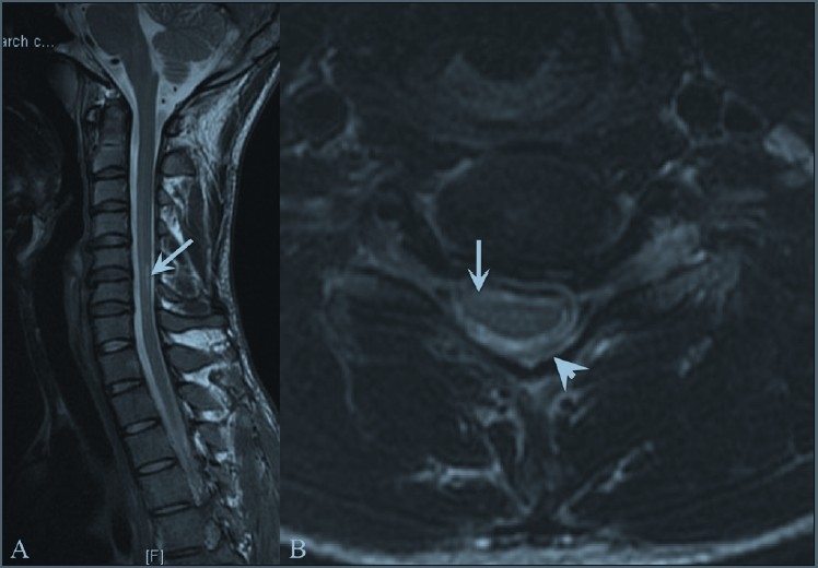
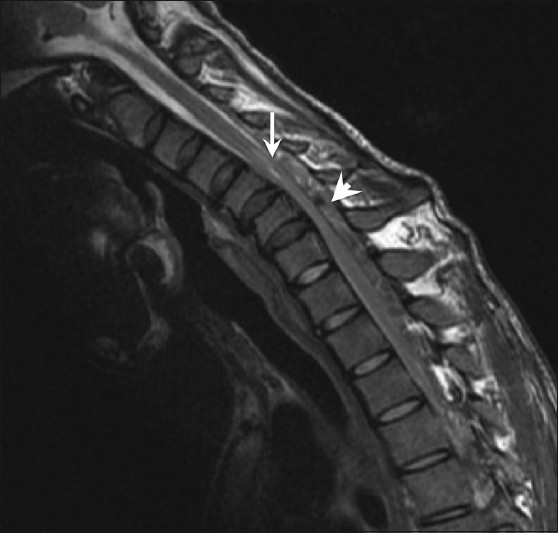
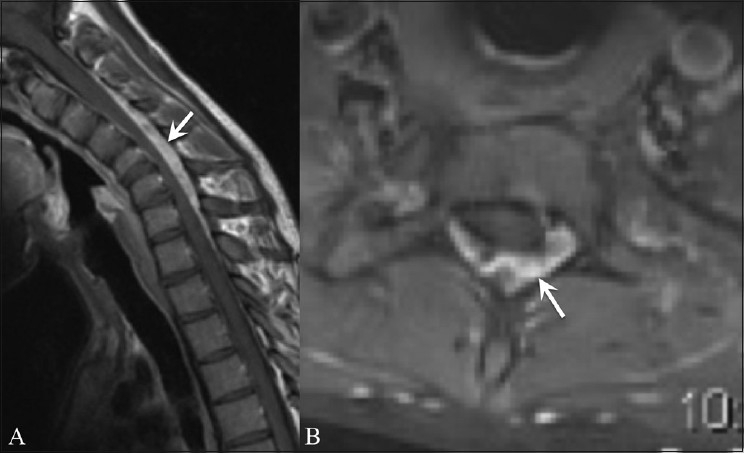
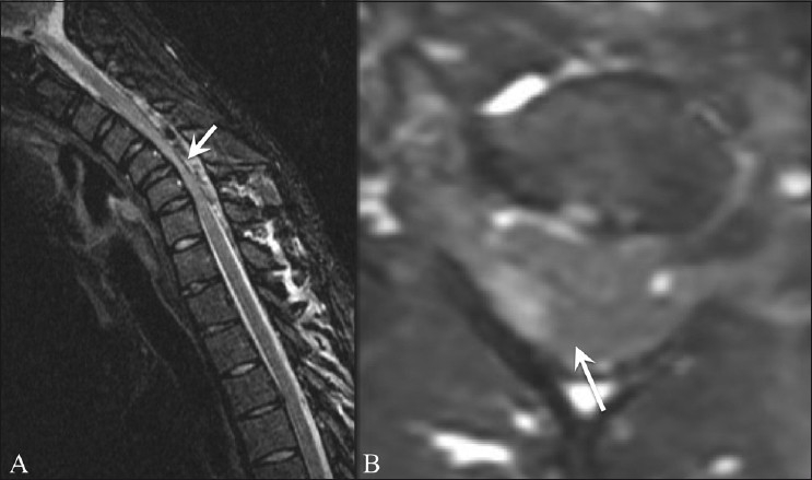
The objective of the study was to study the magnetic resonance imaging (MRI) features of Hirayama disease on a 3 Tesla MRI scanner. Nine patients with clinically suspected Hirayama disease were evaluated with neutral position, flexion, contrast-enhanced MRI and fast imaging employing steady-state acquisition (FIESTA) sequences. The spectrum of MRI features was evaluated and correlated with the clinical and electromyography findings. MRI findings of localized lower cervical cord atrophy (C5-C7), abnormal curvature, asymmetric cord flattening, loss of attachment of the dorsal dural sac and subjacent laminae in the neutral position, anterior displacement of the dorsal dura on flexion and a prominent epidural space were revealed in all patients on conventional MRI as well as with the dynamic 3D-FIESTA sequence. Intramedullary hyperintensity was seen in four patients on conventional MRI and on the 3D-FIESTA sequence. Flow voids were seen in four patients on conventional MRI sequences and in all patients with the 3D-FIESTA sequence. Contrast enhancement of the epidural component was noted in all the five patients with thoracic extensions. The time taken for conventional and contrast-enhanced MRI was about 30-40 min, while that for the 3D-FIESTA sequence was 6 min. Neutral and flexion position MRI and the 3D-FIESTA sequence compliment each other in displaying the spectrum of findings in Hirayama disease. A flexion study should form an essential part of the screening protocol in patients with suspected Hirayama disease. Newer sequences such as the 3D-FIESTA may help in reducing imaging time and obviating the need for contrast.
Keywords: 3D-FIESTA sequence; Flexion MR; Hirayama disease.
Conflict of interest statement
Figures
References
-
- Hirayama K, Tokumaru Y. Cervical dural sac and spinal cord in juvenile muscular atrophy of distal upper extremity. Neurology. 2000;54:1922–6. - PubMed
-
- Hirayama K. Nonprogressive juvenile spinal muscular atrophy of the distal upper limb. In: De Jong JM, editor. Handbook of Clinical Neurology. Amsterdam, Netherlands: Elsevier; 1991. pp. 107–20.
-
- Kikuchi S, Tshiro K, Kitagawa K, Iwasaki Y, Abe H. A mechanism of juvenile muscular atrophy localized in the hand and forearm; Flexion myelopathy with tight dural canal in flexion. Clin Neurol. 1987;27:412–9. - PubMed
-
- Shinomiya K, Sato T, Spengler DM, Dawson J. Isolated muscle atrophy of the of the distal upper extremity in cervical spinal cord compressive disorders. J Spinal Disord. 1995;8:311–6. - PubMed
LinkOut - more resources
Full Text Sources
Miscellaneous