

Ultrasound-guided omental biopsy: Review of 173 patients
- PMID: 21423910
- PMCID: PMC3056632
- DOI: 10.4103/0971-3026.73533
Ultrasound-guided omental biopsy: Review of 173 patients
Abstract
Background: Omental biopsy has conventionally been performed using a surgical approach. USG-guided omental biopsy is a safe and effective alternative. The purpose of this study was to assess the utility of USG guidance for biopsy of the greater omentum.
Study design: Retrospective study.
Materials and methods: We retrospectively reviewed all omental biopsies performed under USG guidance from April 2006 to March 2010 in a tertiary care hospital.
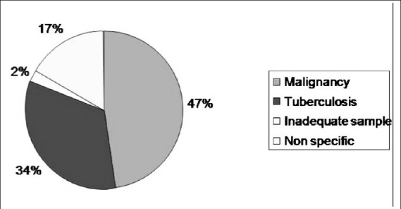
Results: One hundred and seventy-three patients were included. Out of these, 82 (47%) patients were diagnosed to have malignancies, 58 (34%) patients had granulomatous inflammation either suggestive of or consistent with tuberculosis, 29 (17%) patients were diagnosed to have inconclusive biopsy results, and 4 (2%) patients had an inadequate sample for histopathological examination. There were no major procedure-related complications.
Conclusion: USG-guided biopsy of the omentum is a safe and effective procedure. A thickened omentum can serve as an easily accessible site for biopsy, especially in patients who have ascites of unknown etiology and in those with a history of previous malignancy.
Keywords: Biopsy; greater omentum; ultrasound.
Conflict of interest statement
Figures

Similar articles
-
Ultrasound-guided biopsy of greater omentum: an effective method to trace the origin of unclear ascites.Eur J Radiol. 2009 May;70(2):331-5. doi: 10.1016/j.ejrad.2008.01.036. Epub 2008 Mar 6. Eur J Radiol. 2009. PMID: 18328658
-
Role of Ultrasound-Guided Fine-Needle Aspiration Cytology of Omentum in Diagnosis of Abdominal Tuberculosis.Surg Infect (Larchmt). 2019 Jan;20(1):91-94. doi: 10.1089/sur.2018.165. Epub 2018 Nov 27. Surg Infect (Larchmt). 2019. PMID: 30481127
-
Percutaneous omental biopsy: efficacy and complications.Abdom Radiol (NY). 2017 May;42(5):1566-1570. doi: 10.1007/s00261-017-1043-5. Abdom Radiol (NY). 2017. PMID: 28097388
-
Predictive value of omental thickness on ultrasonography for diagnosis of unexplained ascites, an Egyptian centre study.Asian J Surg. 2020 Jan;43(1):13-19. doi: 10.1016/j.asjsur.2019.03.004. Epub 2019 Mar 22. Asian J Surg. 2020. PMID: 30910377 Review.
-
Diagnosis of abdominal tuberculosis: experience from 11 cases and review of the literature.World J Gastroenterol. 2004 Dec 15;10(24):3647-9. doi: 10.3748/wjg.v10.i24.3647. World J Gastroenterol. 2004. PMID: 15534923 Free PMC article. Review.
Cited by
-
Ultrasound Elastography for Differentiating Benign from Malignant Thickened Greater Omentum.Eur Radiol. 2016 Jul;26(7):2337-43. doi: 10.1007/s00330-015-4037-0. Epub 2015 Sep 29. Eur Radiol. 2016. PMID: 26420499
-
Factors influencing diagnostic yield in ultrasound-guided omental biopsies: insights from a retrospective study.Abdom Radiol (NY). 2025 Aug;50(8):3635-3646. doi: 10.1007/s00261-025-04797-z. Epub 2025 Jan 25. Abdom Radiol (NY). 2025. PMID: 39862287
-
Comparative Study of Ultrasound-guided Percutaneous Omental Biopsy in Cirrhotics and Noncirrhotics.J Clin Exp Hepatol. 2020 May-Jun;10(3):194-200. doi: 10.1016/j.jceh.2019.10.003. Epub 2019 Oct 31. J Clin Exp Hepatol. 2020. PMID: 32405175 Free PMC article.
-
Endoscopic Ultrasound (EUS) Guided Fine Needle Aspiration: A New Modality to Diagnose Peritoneal Tuberculosis in Presence of Decompensated Cirrhosis-A Case Series and Review of Literature.J Clin Exp Hepatol. 2018 Jun;8(2):205-209. doi: 10.1016/j.jceh.2017.09.007. Epub 2017 Oct 7. J Clin Exp Hepatol. 2018. PMID: 29892185 Free PMC article.
References
-
- Yoo E, Kim JH, Kim MJ, Yu JS, Chung JJ, Yoo HS, et al. Greater and lesser omenta: Normal anatomy and pathologic processes. Radiographics. 2007;27:707–20. - PubMed
-
- Souza FF, Mortelé KJ, Cibas ES, Erturk SM, Silverman SG. Predictive Value of percutaneous imaging-guided biopsy of peritoneal and omental masses: Results in 111 Patients. AJR Am J Roentgenol. 2009;192:131–6. - PubMed
-
- Sompayrac SW, Mindelzun RE, Silverman PM, Sze R. The greater omentum. AJR Am J Roentgenol. 1997;168:683–7. - PubMed
-
- Ho LM, Thomas J, Fine SA, Paulson EK. Usefulness of sonographic guidance during percutaneous biopsy of mesenteric masses. AJR Am J Roentgenol. 2003;180:1563–6. - PubMed
-
- Spencer JA, Swift SE, Wilkinson N, Boon AP, Lane G, Perren TJ. Peritoneal carcinomatosis: image-guided peritoneal core biopsy for tumor type and patient care. Radiology. 2001;221:173–7. - PubMed
LinkOut - more resources
Full Text Sources
Miscellaneous