

Case Reports
doi: 10.4103/0971-3026.73532.
Disseminated cysticercosis with pulmonary and cardiac involvement
Affiliations
- PMID: 21423911
- PMCID: PMC3056633
- DOI: 10.4103/0971-3026.73532
Item in Clipboard
Case Reports
Disseminated cysticercosis with pulmonary and cardiac involvement
Indian J Radiol Imaging.
2010 Nov.
Abstract
Pulmonary and cardiac involvement by cysticercosis is extremely rare, and is usually asymptomatic. We report the case of a 19-year-old boy who presented with a history of headache and vomiting and was found to have disseminated cysticercosis with pulmonary and cardiac involvement; the emphasis is on the rare occurrence of pulmonary, cardiac, pancreatic, intraocular, and extradural spinal canal involvement in the same patient. This case demonstrates the extent to which cysticercosis can be disseminated.
Keywords: Cardiac; disseminated cysticercosis; intraocular; pulmonary.
Conflict of interest statement
Figures

T2W axial MRI of the brain shows multiple cystic lesions with hypointense eccentric nodules, in both cerebral hemispheres, the midbrain, the cerebellum, and the extraocular muscles
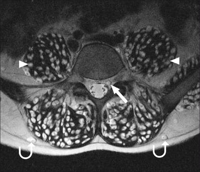
T2W axial MRI of the lumbar spine shows cysticercus cysts in the extradural spinal space (arrow), in addition to muscle (arrowhead) and subcutaneous (curved arrow) involvement
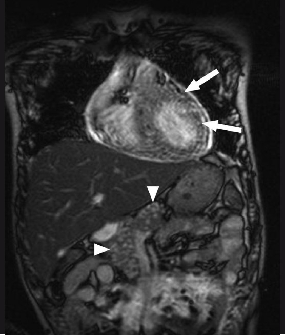
Balanced gradient MRI coronal image of the chest and upper abdomen shows hyperintense nodular lesions in the cardiac muscles (arrows) and pancreas (arrowhead)
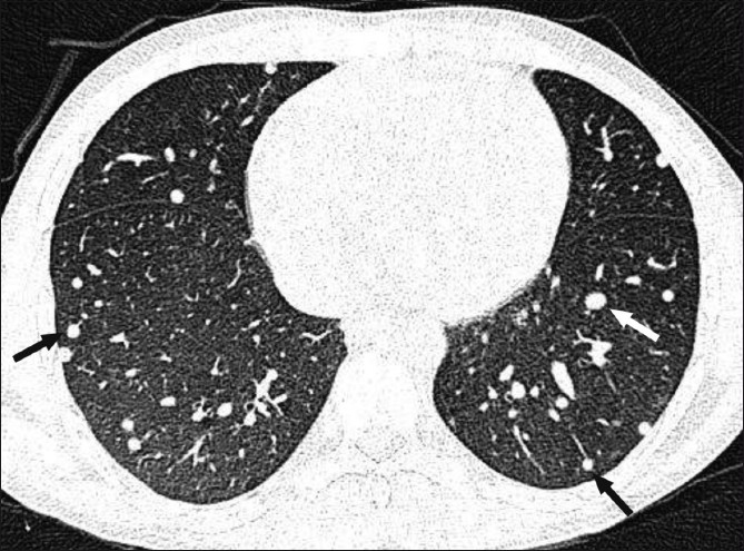
HRCT of the lungs shows multiple randomly distributed nodules (arrows) of varying sizes
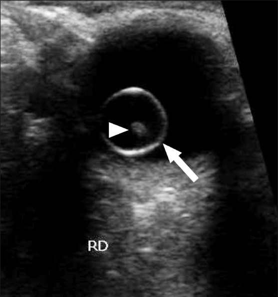
Transverse B-scan of the right eye shows a large intravitreal cyst (arrow) with a tiny hyperechoic scolex (arrowhead) within it
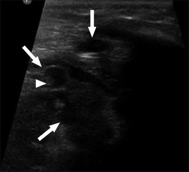
USG of the heart shows multiple cystic anechoic lesions (arrow) in the myocardium with a tiny hyperechoic scolex (arrowhead) within one of the cysts
Similar articles
-
Asymptomatic disseminated cysticercosis.J Clin Diagn Res. 2013 Aug;7(8):1761-3. doi: 10.7860/JCDR/2013/5465.3269. Epub 2013 Aug 1. J Clin Diagn Res. 2013. PMID: 24086907 Free PMC article.
-
Simultaneous intraocular and bilateral extraocular muscle involvement in a case of disseminated cysticercosis.Int Ophthalmol. 2005 Feb-Apr;26(1-2):35-7. doi: 10.1007/s10792-005-8248-2. Epub 2006 Jun 15. Int Ophthalmol. 2005. PMID: 16779570
-
A rare case of disseminated cysticercosis: case report and literature review.Indian J Med Microbiol. 2011 Apr-Jun;29(2):180-3. doi: 10.4103/0255-0857.81787. Indian J Med Microbiol. 2011. PMID: 21654117 Review.
-
[Myocardial localization of a disseminated cysticercosis. Echocardiographic diagnosis of a case].Arch Mal Coeur Vaiss. 2002 Jun;95(6):606-8. Arch Mal Coeur Vaiss. 2002. PMID: 12138821 French.
-
Spinal extradural cysticercosis: a case report.Spinal Cord. 1998 Apr;36(4):285-7. doi: 10.1038/sj.sc.3100524. Spinal Cord. 1998. PMID: 9589530 Review.
Cited by
-
Isolated pancreatic cysticercal cyst presenting as a diagnostic challenge: diagnosis and treatment review.BMJ Case Rep. 2015 Jul 9;2015:bcr2015210774. doi: 10.1136/bcr-2015-210774. BMJ Case Rep. 2015. PMID: 26160552 Free PMC article.
-
Challenges in the Diagnosis of Taenia solium Cysticercosis and Taeniosis in Medical and Veterinary Settings in Selected Regions of Tanzania: A Cross-Sectional Study.Vet Med Int. 2022 Jun 30;2022:7472051. doi: 10.1155/2022/7472051. eCollection 2022. Vet Med Int. 2022. PMID: 35815231 Free PMC article.
-
Disseminated neurocysticercosis presenting as affective mood disorder with chronic tension type headache.J Neurosci Rural Pract. 2013 Oct;4(4):461-3. doi: 10.4103/0976-3147.120212. J Neurosci Rural Pract. 2013. PMID: 24347961 Free PMC article.
-
Isolated cardiac cysticercosis of the right ventricle.Indian J Thorac Cardiovasc Surg. 2024 Jan;40(1):99-102. doi: 10.1007/s12055-023-01617-1. Epub 2023 Nov 8. Indian J Thorac Cardiovasc Surg. 2024. PMID: 38125329 Free PMC article.
-
A rare case of disseminated cysticercosis.Trop Parasitol. 2012 Jul;2(2):138-41. doi: 10.4103/2229-5070.105183. Trop Parasitol. 2012. PMID: 23767025 Free PMC article.
References
-
- Nutman TB, Weller PF. Cestodes. In: Fauci AS, editor. Harrisons Principles of internal medicine. 14th ed. USA: McGraw-Hill; 1998. p. 1225.
-
- Mamere AE, Muglia VF, Belucci AD, Carlos dos Santos A, Trad CS, Takayanagui OM. Disseminated cysticercosis with pulmonary involvement. J Thorac Imaging. 2004;19:109–11. - PubMed
-
- Murray PR, Rosenthal KS, Pfaller MA. Cestodes. In: Murray PR, editor. Medical Microbiology. 5th ed. USA: Elsevier Mosby; 2005. p. 908.
-
- King CH. Cestodes (Tapeworms) In: Mandell GL, editor. Principles and practice of infectious diseases. 6th ed. Philadelphia: Elsevier Churchill Livingstone; 2005. p. 3289.
Publication types
LinkOut - more resources
Full Text Sources