

Impact of analyzing fewer image frames per segment during offline volumetric radiofrequency-based intravascular ultrasound measurements of target lesions prior to percutaneous coronary interventions
- PMID: 21424153
- PMCID: PMC3326371
- DOI: 10.1007/s10554-011-9843-0
Impact of analyzing fewer image frames per segment during offline volumetric radiofrequency-based intravascular ultrasound measurements of target lesions prior to percutaneous coronary interventions
Abstract
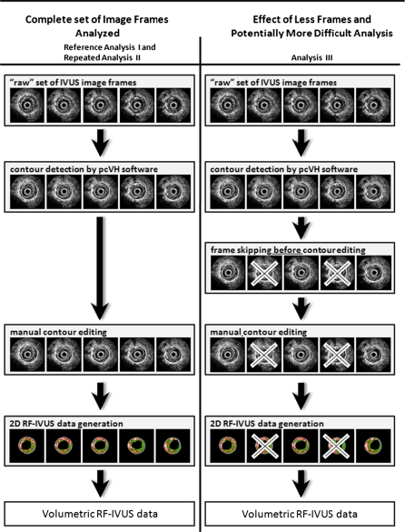
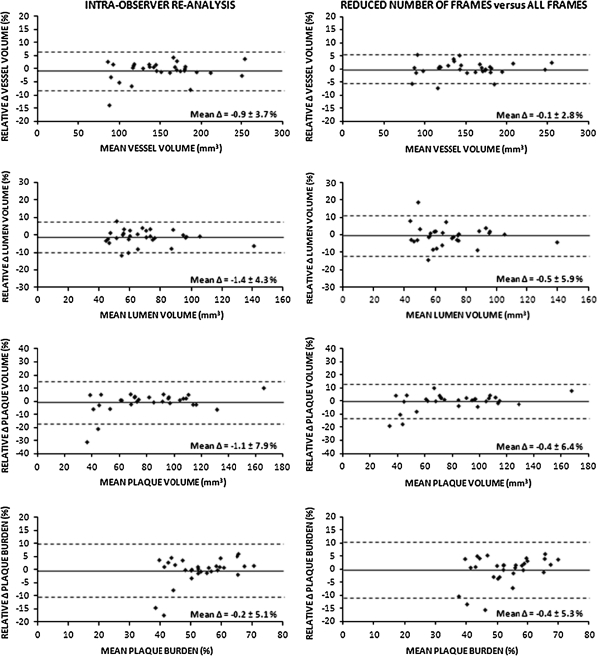
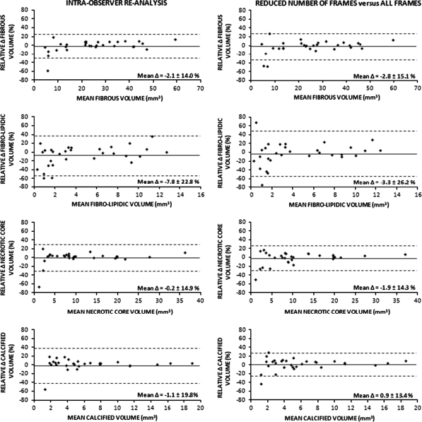
In the present study, we evaluated the impact of a 50% reduction in number of image frames (every second frame) on the analysis time and variability of offline volumetric radiofrequency-based intravascular ultrasound (RF-IVUS) measurements in target lesions prior to percutaneous coronary interventions (PCI). Volumetric RF-IVUS data of vessel geometry and plaque composition are generally obtained by a semi-automated analysis process that includes time-consuming manual contour editing. A reduction in the number of frames used for volumetric analysis may speed up the analysis, but could increase measurement variability. We repeatedly performed offline volumetric analyses in RF-IVUS image sets of 20 mm-long coronary segments that contained 30 de novo lesions prior to PCI. A 50% reduction in frames decreased the analysis time significantly (from 57.5 ± 7.3 to 35.7 ± 3.7 min; P < 0.0001) while geometric and compositional RF-IVUS measurements did not differ significantly from measurements obtained from all frames. The variability between measurements on the reduced number of frames versus all frames was comparable to the intra-observer measurement variability. In target lesions prior to PCI, offline volumetric RF-IVUS analyses can be performed using a reduced number of image frames (every second frame). This reduces the time of analysis without substantially increasing measurement variability.
Figures
References
-
- Bruining N, Verheye S, Knaapen M, Somers P, Roelandt JR, Regar E, Heller I, de Winter S, Ligthart J, Van Langenhove G, de Feijter PJ, Serruys PW, Hamers R. Three-dimensional and quantitative analysis of atherosclerotic plaque composition by automated differential echogenicity. Catheter Cardiovasc Interv. 2007;70(7):968–978. doi: 10.1002/ccd.21310. - DOI - PubMed
-
- Di Mario C, Gorge G, Peters R, Kearney P, Pinto F, Hausmann D, von Birgelen C, Colombo A, Mudra H, Roelandt J, Erbel R (1998) Clinical application and image interpretation in intracoronary ultrasound. Study group on intracoronary imaging of the working group of coronary circulation and of the subgroup on intravascular ultrasound of the working group of echocardiography of the European society of cardiology. Eur Heart J 19(2):207–229 - PubMed
-
- Mintz GS, Nissen SE, Anderson WD, Bailey SR, Erbel R, Fitzgerald PJ, Pinto FJ, Rosenfield K, Siegel RJ, Tuzcu EM, Yock PG (2001) American college of cardiology clinical expert consensus document on standards for acquisition, measurement and reporting of intravascular ultrasound studies (IVUS). A report of the American college of cardiology task force on clinical expert consensus documents. J Am Coll Cardiol 37(5):1478–1492 - PubMed
-
- Garcia-Garcia HM, Mintz GS, Lerman A, Vince DG, Margolis MP, van Es GA, Morel MA, Nair A, Virmani R, Burke AP, Stone GW, Serruys PW. Tissue characterisation using intravascular radiofrequency data analysis: recommendations for asquisition, analysis, interpretation and reporting. Eurointervention. 2009;5(2):177–189. doi: 10.4244/EIJV5I2A29. - DOI - PubMed
MeSH terms
LinkOut - more resources
Full Text Sources
Medical
Miscellaneous
