. 2012 Feb;44(1):183-96.
doi: 10.1007/s11255-011-9932-y. Epub 2011 Mar 19.

Histological heterogeneity of glomerular segmental lesions in focal segmental glomerulosclerosis

Affiliations

Histological heterogeneity of glomerular segmental lesions in focal segmental glomerulosclerosis

Sekiko Taneda et al. Int Urol Nephrol. 2012 Feb.

Abstract

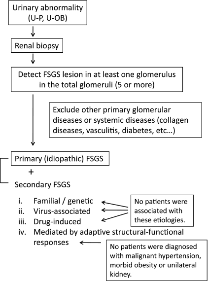
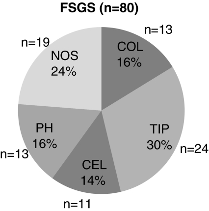
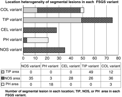
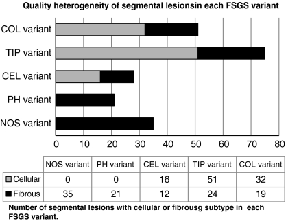
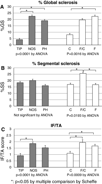
Focal segmental glomerulosclerosis (FSGS) involves considerable histological heterogeneity in terms of location and quality of the glomerular segmental lesions. The present study investigated the heterogeneity of segmental lesions in each variant of FSGS, determined by the Columbia classification, and its clinical relevance. All glomerular segmental lesions of 80 cases of primary FSGS were evaluated histologically based on location [tip (TIP), perihilar (PH), or not otherwise specified (NOS)], and quality (cellular or fibrous). Among the 1,299 glomeruli of the 80 biopsy specimens, 210 glomeruli (16.2%) had segmental lesions, comprising 57 (27%) cellular TIP, 4 (2%) fibrous TIP, 42 (20%) cellular NOS, 86 (41%) fibrous NOS, and 21 (10%) fibrous PH lesions. Each case was also classified into one of the five histological variants of the Columbia classification: collapsing (COL), TIP, cellular (CEL), PH, or NOS. Overlap of segmental lesions in different location categories was seen in the COL, TIP, and PH variants, and heterogeneity of quality was apparent in the COL and CEL variants. Histological findings of the CEL variant (endocapillary hypercellularity) were observed in nine of the 13 COL variants. Both location and quality correlated with disease duration, degree of proteinuria, and histological severity of global glomerular sclerosis and tubulo-interstitial lesions. These results demonstrated the histological heterogeneity of glomerular segmental lesions in all variants of the Columbia classification, except NOS. However, the fidelity of location and dominance of histological features were generally conserved in the TIP and PH variants. The COL and CEL variants warrant further investigation because of their overlapping histological findings and apparent histological heterogeneity in the glomerular segmental lesions.


Figures

Fig. 1
Fig. 1
Screening program for diagnosis and etiologies of FSGS. FSGS was diagnosed based on the presence of glomerular segmental lesions in at least one glomerulus. Patients diagnosed with other primary glomerular diseases or systemic disease, such as collagen diseases, vasculitis, or diabetes, were excluded. No patient was identified with a cause for secondary FSGS. Thus, the subjects of this study were diagnosed mostly with primary (idiopathic) FSGS
Fig. 2
Fig. 2
Histological variants according to the Columbia classification. The 80 cases of FSGS were classified according to the Columbia classification as follows: COL, 13 cases (16.2%), TIP, 24 cases (30.0%), CEL, 11 cases (13.8%), PH, 13 cases (16.2%), and NOS, 19 cases (23.8%)
Fig. 3
Fig. 3
Typical glomerular findings of segmental lesions regarding location and histological features. a TIP-located lesion with cellular histology (TIP-cellular). There is segmental accumulation of endocapillary foam cells involving the peripheral glomerular segment at the tubular pole. Podocytes overlying the segmental lesion are hypertrophic and hyperplastic. The histological variant of this glomerulus corresponds to TIP in the Columbia classification (silver methenamine stain, ×100). b TIP-located lesion with fibrous histology (TIP-fibrous). The glomerular tufts are collapsed with segmental accumulation of extracellular matrix involving the peripheral glomerular segment at the tubular pole. Podocytes overlying the segmental lesion are slightly proliferative. The histological variant of this glomerulus also corresponds to TIP in the Columbia classification. As a TIP-cellular lesion co-existed in the same specimen, the histological category of this case was deemed TIP-fibrocellular (F/C) (PAS stain, ×100). c Extensive segmental lesion in the NOS area with cellular histology (NOS-cellular). The glomerulus shows extensive collapse and obliteration of the glomerular tufts, accompanying prominent hypertrophy and hyperplasia of the overlying glomerular epithelial cells. This case is categorized as NOS-cellular and classified as a COL variant in the Columbia classification (silver methenamine stain, ×100). d Segmental lesion in the NOS area with active endocapillary cellular histology (NOS-cellular). Endocapillary accumulation of foam cells and leukocytes occluding the glomerular capillary lumina is regarded as an active histological feature of glomerular segmental lesions. This case is categorized as NOS-cellular and classified as a CEL variant in the Columbia classification. In addition to endocapillary proliferation, collapsing glomerular tufts may also be observed in some areas of the segmental lesion, making it difficult to differentiate between the COL and CEL variants (silver methenamine stain, ×100). e NOS-located lesion with a predominantly fibrous appearance (NOS-fibrous). Extensive segmental sclerosis is observed involving the NOS area, but at neither the vascular nor tubular pole. No cellular proliferation is seen in the segmental lesion. The case is categorized as NOS-fibrous and classified as NOS in the Columbia classification (PAS stain, ×100). f PH-located lesion showing segmental occlusion of the glomerular capillaries by fibrous matrix accumulation and hyalinosis around the glomerular hilum (PH-fibrous). Neither epithelial hyperplasia nor endothelial proliferation is seen in this glomerulus. This case is categorized as PH-fibrous and classified as the PH variant in the Columbia classification (PAS stain, ×100)
Fig. 4
Fig. 4
Composition of location and quality factors in all segmental lesions (n = 210). The 210 glomerular segmental lesions consisted of 57 (27%) cellular TIP, four (2%) fibrous TIP, 42 (20%) cellular NOS, 86 (41%) fibrous NOS, and 21 (10%) fibrous PH lesions. The majority of segmental lesions in the TIP area were cellular, whereas all 21 segmental lesions in the PH area were fibrous. The segmental lesions in the NOS area were mixed cellular (33.1%) and fibrous (66.9%)
Fig. 5
Fig. 5
Location heterogeneity of segmental lesions in each histological variant. Heterogeneity in terms of location was present of the segmental lesions. In the COL variant, segmental lesions were located in the TIP (12/51, 23.5%), NOS (36/51, 70.6%), and PH areas (3/51, 5.9%). In the TIP variant, segmental lesions were located in the TIP (49/75, 65.3%) and NOS areas (26/75, 34.7%), but not in the PH area. All segmental lesions in the CEL variant were located in the NOS area (28/28, 100%). Most of the segmental lesions in the PH variant were located in the PH area (18/21, 85.7%), with three located in the NOS area (3/21, 14.3%). All segmental lesions in the NOS variant were located in the NOS area (35/35, 100%). Gray bars, TIP area; dashed bars, NOS area; dotted bars, PH area
Fig. 6
Fig. 6
Quality heterogeneity of segmental lesions in each histological variant. The quality composition, cellular (white bars) and fibrous (black bars) segmental lesions, in each variant is shown. Among the 210 segmental lesions, 99/210 (47.1%) were cellular and 111/210 (52.9%) were fibrous. In the COL variant, 32/51 (62.7%) were cellular and 19/51 (37.3%) were fibrous. The cellular subtype was also predominant in the TIP (51/75, 68%) and CEL variants (16/28, 57.1%). In contrast, all segmental lesions were fibrous in the NOS and PH variants. Gray bars, cellular; black bars, fibrous
Fig. 7
Fig. 7
Overlap of COL and CEL lesion histological findings. The glomerulus showed the coexistence of segmental tuft collapse (arrow head) and endocapillary hypercellularity with intracapillary foam cells (arrow). Overlap between the CEL lesion in the COL variant sometimes made it difficult to determine the variant. The CEL lesion was present in nine of 13 cases with COL variants in this study (silver methenamine stain, ×200)
Fig. 8
Fig. 8
Correlation between clinical parameters and dominant location/quality factors of segmental lesions. a Interval between onset and biopsy (months). b Urinary protein excretion at biopsy (g/day). c Serum creatinine (sCr) level at final observation (mg/dl). Both the location and quality of lesions were significantly correlated with disease duration and proteinuria at biopsy. The outcome, as reflected by the sCr at the end of follow-up, was significantly correlated with location, but not with quality of lesions
Fig. 9
Fig. 9
Correlation between histological parameters and dominant location/quality factors of segmental lesions. a Globally sclerotic glomeruli as a percentage of the total number of glomeruli (%GS). b Segmentally sclerotic glomeruli as a percentage of the total number of glomeruli (%SS). c Score of interstitial fibrosis and tubular atrophy (IF/TA score). Both the location and quality of lesions were significantly correlated with %GS and the IF/TA score. Quality was correlated only weakly and location not at all with %SS, meaning that the percentage of segmentally sclerotic glomeruli was similar across location categories and quality subtypes

References

    1. Schwartz MM, Evans J, Bain R, et al. Focal segmental glomerulosclerosis: prognostic implications of the cellular lesion. J Am Soc Nephrol. 1999;10:1900–1907. - PubMed
    1. D’Agati VD. The spectrum of focal segmental glomerulosclerosis: new insights. Curr Opin Nephrol Hypertens. 2008;17:271–281. doi: 10.1097/MNH.0b013e3282f94a96. - DOI - PubMed
    1. D’Agati VD, Fogo AB, Bruijn JA, et al. Pathologic classification of focal segmental glomerulosclerosis: a working proposal. Am J Kidney Dis. 2004;43:368–382. doi: 10.1053/j.ajkd.2003.10.024. - DOI - PubMed
    1. Stokes MB, Markowitz GS, Lin J, et al. Glomerular tip lesion: a distinct entity within the minimal change disease/focal segmental glomerulosclerosis spectrum. Kidney Int. 2004;65:1690–1702. doi: 10.1111/j.1523-1755.2004.00563.x. - DOI - PubMed
    1. Thomas DB, Franceschini N, Hogan SL, et al. Clinical and pathologic characteristics of focal segmental glomerulosclerosis pathologic variants. Kidney Int. 2006;69:920–926. doi: 10.1038/sj.ki.5000160. - DOI - PubMed

MeSH terms

LinkOut - more resources