

Intradural disc herniation at L5 level mimicking an intradural spinal tumor
- PMID: 21424915
- PMCID: PMC3111494
- DOI: 10.1007/s00586-011-1772-z
Intradural disc herniation at L5 level mimicking an intradural spinal tumor
Abstract
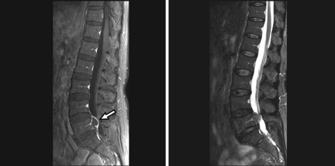
Intradural lumbar disc herniation is a rare complication of disc disease. The reason for the tearing of the dura matter by a herniated disc is not clearly known. Intradural disc herniations usually occur at the disc levels and are often seen at L4-L5 level but have also been reported at other intervertebral disc levels. However, intradural disc herniation at mid-vertebral levels is rare in the literature and mimics an intradural extramedullary spinal tumor lesion in radiological evaluation. Although magnetic resonance imaging (MRI) with gadolinium is useful in the diagnosis of this condition, preoperative correct diagnosis is usually difficult and the definitive diagnosis must be made during surgery. We describe here a 50-year-old female patient who presented with pain in the lower back for 6 months and a sudden exacerbation of the pain that spread to the left leg as well as numbness in both legs for 2 weeks. MRI demonstrated an intradural mass at the level of L5. Laminectomy was performed, and subsequently durotomy was also performed. An intradural disc fragment was found and completely removed. The patient recovered fully in 3 months. Intradural lumbar disc herniation must be considered in the differential diagnosis of mass lesions in the spinal canal.
Figures


References
-
- Dandy WE. Serious complications of ruptured intervertebral discs. JAMA. 1942;11:474–477.
-
- Epstein NE, Syrquin MS, Epstein JA, Decker RE. Intradural disc herniations in the cervical, thoracic, and lumbar spine: report of three cases and review of the literature. J Spinal Disord. 1990;3:396–403. - PubMed
Publication types
MeSH terms
LinkOut - more resources
Full Text Sources
Medical
