Review
. 2011 Apr;8(2):152-67.
doi: 10.1007/s13311-011-0026-4.

Antioxidant therapies for acute spinal cord injury

Affiliations
Review

Antioxidant therapies for acute spinal cord injury

Edward D Hall. Neurotherapeutics. 2011 Apr.

Abstract

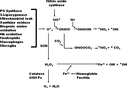
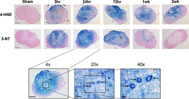
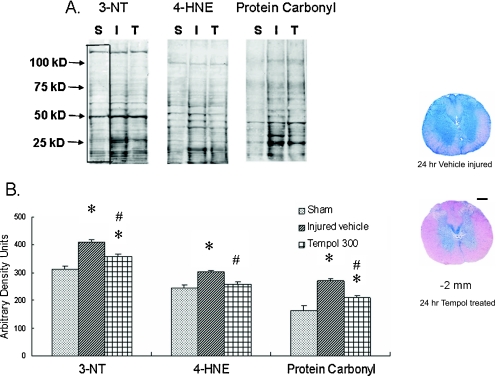
One of the most investigated molecular mechanisms involved in the secondary pathophysiology of acute spinal cord injury (SCI) is free radical-induced, iron-catalyzed lipid peroxidation (LP) and protein oxidative/nitrative damage to spinal neurons, glia, and microvascular cells. The reactive nitrogen species peroxynitrite and its highly reactive free radicals are key initiators of LP and protein nitration in the injured spinal cord, the biochemistry, and pathophysiology of which are first of all reviewed in this article. This is followed by a presentation of the antioxidant mechanistic approaches and pharmacological compounds that have been shown to have neuroprotective properties in preclinical SCI models. Two of these, which act by inhibition of LP, are high-dose treatment with the glucocorticoid steroid methylprednisolone (MP) and the nonglucocorticoid 21-aminosteroid tirilazad, have been demonstrated in the multicenter NASCIS clinical trials to produce at least a modest improvement in neurological recovery when administered within the first 8 hours after SCI. Although these results have provided considerable validation of oxidative damage as a clinically practical neuroprotective target, there is a need for the discovery of safer and more effective antioxidant compounds for acute SCI.


Figures

FIG. 1
FIG. 1
Biochemistry of oxygen radical formation via the peroxynitrite pathway and the dismutation/Fenton reaction pathway. Nomenclature: CO2 = carbon dioxide; •CO3 = carbonate radical; Fe2+ = ferrous iron; Fe3+ = ferric iron; GSH PX = glutathione peroxidase; H+ = proton; H2O2 = hydrogen peroxide; Hb = hemoglobin; NO• = nitric oxide or nitrogen monoxide; •NO2 = nitrogen dioxide; O2-. = superoxide radical; •OH = hydroxyl radical; ONOO- = peroxynitrite anion; ONOOCO2- = nitrosoperoxocarbonate; ONOOH = peroxynitrous acid; PG = prostaglandin; SOD = superoxide dismutase.
FIG. 2
FIG. 2
(a) Representative examples showing the post-traumatic time course of oxidative damage as revealed by 3-nitrotyrosine (3-NT) and 4-hydroxynonenal (4-HNE) immunostaining. Images are representative coronal sections at the epicenter (T10) at 24 and 72 h, and at 1 and 2 weeks post-injury. Adjacent sections from the same animal were stained for 3-NT and 4-HNE. All sections are counterstained with nuclear fast red. Sham animal showed minimal staining for both markers. By 3-h post injury, staining was substantially elevated, encompassing all of the gray matter and extending into the white matter. It peaked for 3-NT and 4-HNE around 24- to 72-h postinjury with only a small rim of white matter remaining unstained. By 1 week, 3-NT staining had nearly disappeared, whereas the elevation of 4-HNE persisted throughout the gray matter out to at least 2-week post injury. Scale bar = 500 μm. (b) Higher power views of 3-NT oxidative damage in the injured spinal cord in the 24-h post-injury spinal cross section at 1-mm caudal to the epicenter, which shows intense oxidative damage staining of gray matter microvessels (arrows), as well as surrounding parenchymal elements, the same 24-h 3-NT cross section from (a). A collage of FIGS. 1 and 5, and for more detail see Carrico et al. [22], reproduced with permission.
FIG. 3
FIG. 3
(a) Representative Western blots shows staining patterns of 3-nitrotyrosine (3-NT), 4-hydroxynonenal (4-HNE), and protein carbonyls. S, I, T = sham, vehicle, and tempol-treated groups (300 mg/kg, i.p.), respectively. Compared to the sham group, more intense staining patterns are observed in vehicle groups and tempol treatment attenuated the immunoreactivity. (b) Densitometric quantification of 3-NT, 4-HNE, and protein carbonyls (across various molecular weight ranges, included in the black box in each lane) showed significant injury-induced increases in peroxynitrite (PN) formation, lipid peroxidation and protein oxidation, which were each decreased by the PN-derived radical scavenger tempol compared to the saline-treated injured group (values = mean ± standard error; *p < 0.05 vs sham; #p < 0.05 vs vehicle; n = 6/group). (c) Representative spinal cord cross sections 2 mm from the epicenter of the contusion injury from rats and treated with saline vehicle or tempol are shown Scale bar = 0.5 mm. This figure is a collage of parts of FIGS. 2 and 3, and for more detail see Xiong et al. [105], reproduced with permission.

References

    1. Hall ED, Vaishnav RA, Mustafa AG. Antioxidant therapies for traumatic brain injury. Neurotherapeutics. 2010;7:51–61. - PMC - PubMed
    1. Halliwell B, Gutteridge J. Free Radicals in Biology and Medicine, 3 rd ed. Oxford University Press, 2008.
    1. Zaleska MM, Floyd RA. Regional lipid peroxidation in rat brain in vitro: possible role of endogenous iron. Neurochem Res. 1985;10:397–410. - PubMed
    1. Sadrzadeh SM, Graf E, Panter SS, Hallaway PE, Eaton JW. Hemoglobin: a biologic fenton reagent. J Biol Chem. 1984;259:14354–14356. - PubMed
    1. Sadrzadeh SM, Eaton JW. Hemoglobin-mediated oxidant damage to the central nervous system requires endogenous ascorbate. J Clin Invest. 1988;82:1510–1515. - PMC - PubMed

Publication types