. 2011 Apr;69(4):721-34.
doi: 10.1002/ana.22339. Epub 2011 Mar 18.

Interleukin-2/interleukin-2 antibody therapy induces target organ natural killer cells that inhibit central nervous system inflammation

Affiliations

Interleukin-2/interleukin-2 antibody therapy induces target organ natural killer cells that inhibit central nervous system inflammation

Junwei Hao et al. Ann Neurol. 2011 Apr.

Abstract

Objective: The role of natural killer (NK) cells in regulating multiple sclerosis (MS) is not well understood. Additional studies with NK cells might provide insight into the mechanism of action of MS therapies such as daclizumab, an antibody against the interleukin (IL)-2R α-chain, which induces expansion of CD56(bright) NK cells.

Methods: In a relapsing-remitting form of the experimental autoimmune encephalomyelitis (EAE) model of MS induced in SJL mice, we expanded NK cells with IL-2 coupled with an anti-IL-2 monoclonal antibody (mAb) and evaluated the effects of these NK cells on EAE. Further, we investigated the effect of the human version of IL-2/IL-2 mAb on NK cells from MS patients and its effect on central nervous system (CNS) inflammation and pathology in a human-mouse chimera model and assessed the underlying mechanisms.

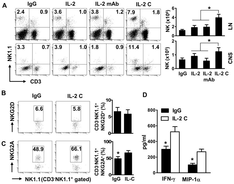
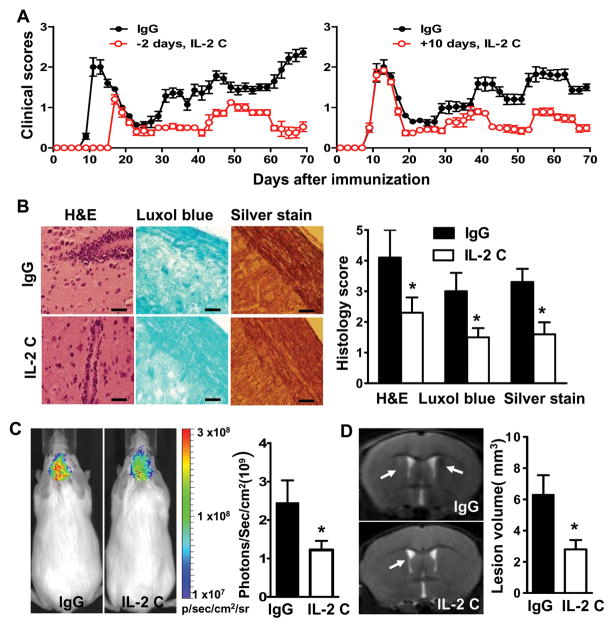
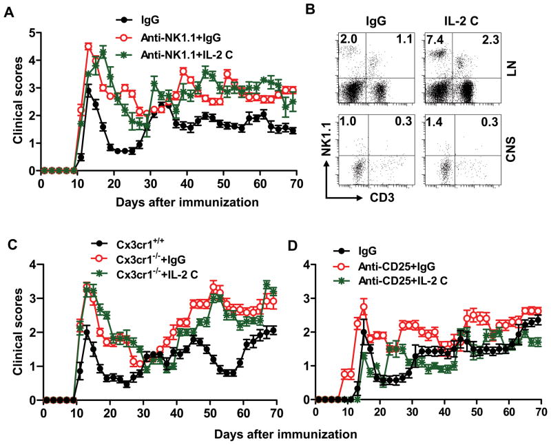
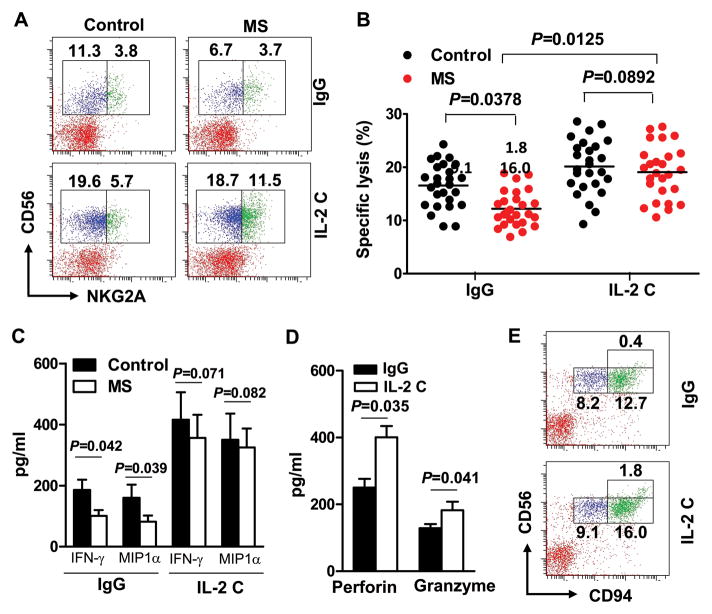
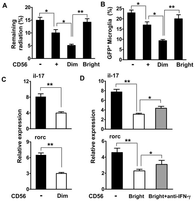
Results: IL-2/IL-2 mAb dramatically expands NK cells both in the peripheral lymphoid organs and in the CNS, and attenuates CNS inflammation and neurological deficits. Disease protection is conferred by CNS-resident NK cells. Importantly, the human version of IL-2/IL-2 mAb restored the defective CD56(+) NK cells from MS patients in a human-mouse chimera model. Both the CD56(bright) and CD56(dim) subpopulations were required to attenuate disease in this model.

Interpretation: These findings unveil the immunotherapeutic potential of NK cells, which can act as critical suppressor cells in target organs of autoimmunity. These results also have implications to better understand the mechanism of action of daclizumab in MS.


Conflict of interest statement

Potential Conflicts of Interest

D. Campagnolo is an employee and stock holder of Biogen Idec. Inc. T. Vollmer received research grants and honoraria from Biogen Idec. Inc. and was involved in clinical trials of daclizumab.

Figures

FIGURE 1
FIGURE 1. IL-2/anti-IL-2 mAb complexes expand NK cells in the periphery and the CNS
After EAE induction with PLP, SJL/J mice were treated with control IgG, IL-2, anti-IL-2 mAb (S4B6), or a combination of IL-2 and anti-IL-2 mAb (IL-2 C) i.p., on the same day of PLP139–151 immunization until termination of the experiment, as described in detail in the M & M. Single cell suspensions were prepared from spleens and CNS on days 10–20 post immunization (p.i.). Frequency and phenotypes of mononuclear cells were analyzed by FACS. (A) Frequency and numbers of NK and NKT cells in the periphery and CNS on gated lymphocytes in corresponding compartments. (B, C) Expression of NKG2A and NKG2D on gated NK1.1+CD3 cells. (D) Lymphocytes isolated from mouse LN tissues were cultured with IgG or IL-2 C for 48–72 hours. Production of IFN-γ and MCP-1α by sorted NK cells was measured by ELISA. The data of one experiment out of two performed is shown, n = 3–4 mice/group. Dot plots are representative of two separate experiments (n=6–18 mice). P values, Student’s t-test, *p<0.05.
FIGURE 2
FIGURE 2. IL-2 complexes inhibit EAE activity and CNS pathology
Mice were immunized with PLP/CFA and treated with IgG or IL-2 complexes (IL-2 C) prior to or after immunization. (A) EAE scores were recorded as mean clinical scores ± SD. Left panel denotes IL-2 C administration before or during EAE induction, n= 18–20 mice/group; right panel IL-2 C administration at day 10 p.i., n=6–8 mice/group. (B) Immunohistochemical analysis of intensity of infiltration, demyelination and axonal loss of spinal cords harvested at days 10–25 p.i. Results are representative of two experiments. (C) In vivo bioluminescence imaging analyses of brains at days 10–25 p.i. using Xenogen IVIS spectrum. Results are expressed as the region of interest (ROI) measurements of brain by Loess regression statistical analyses. (D) Visualization of brain demyelination with 7TMRI. Arrow heads indicate focal lesions located around the lateral ventricle and increased signal intensity on T2-weighted lesions. Pathology and imaging experiments were conducted in groups of mice (n= 4) between 10–25 days post immunization. Data are representative of two independent experiments (mean and s.e.m.). Scale bar, 100 μm. P values for clinical scores were evaluated by Mann-Whitney U test, all other comparisons between groups were analyzed by Student’s t-test for two groups and ANOVA for multiple comparisons; *p<0.05, **p<0.01.
FIGURE 3
FIGURE 3. IL-2 complexes preferentially inhibit myelin-reactive Th17 cells in the CNS
Mice were primed with PLP/CFA and treated with IgG or IL-2 complexes (IL-2 C). Mice were sacrificed at day 10–25 post-immunization (p.i.). Lymph node and CNS cells were isolated. (A) Expansion of CD4+ T cells in the periphery and in the CNS on gated lymphocytes was assessed by CFSE dilution or BrdU incorporation assay, respectively. (B) Lymphoid or CNS cells were re-stimulated with PLP overnight and IFN-γ- and IL-17-expressing CD4+ T cells were measured by intracellular staining. FACS dot plots are representative of four independent experiments (n= 6–15/group). Bar data were accumulated from four independent experiments; P values were calculated by Student’s t-test.
FIGURE 4
FIGURE 4. Effects of IL-2 complexes on EAE are dependent on CNS-resident NK cells
Groups of mice were immunized with PLP/CFA and treated with IgG or IL-2 complexes (IL-2 C) as indicated in M&M. (A) Mice received anti-NK1.1 mAb prior to immunization until termination of the experiment. Clinical features of EAE in these mice were monitored and compared. n=8–12/group. (B) Frequency of NK cells in lymph node and CNS from SJL Cx3cr1−/− mice at days 10–70 p.i. Results are representative of four experiments with 5–12 mice per group each. (C) Clinical features of EAE in SJL Cx3cr1−/− mice that were treated with IgG or IL-2 complexes were monitored and compared. n=10–-12/group. (D) Effects of IL-2 complexes are preserved in PLP/CFA primed mice after CD25+ T cell depletion. n = 8/group. P values for clinical scores were evaluated by Mann-Whitney U test, other comparisons between groups were analyzed by Student’s t-test; *p<0.05, **p<0.01.
FIGURE 5
FIGURE 5. IL-2 complexes restore defective NK cells from MS patients
Blood was drawn from relapsing remitting MS patients or healthy controls. Peripheral blood mononuclear cells (PBMC) were isolated and incubated with IgG or a combination of IL-2 (10 ng) and anti-IL-2 mAb (10 μg/ml) for 48–72 hours. (A) Frequency of CD56 and NKG2A double-positive cells on gated CD3 lymphocytes. Plots are representative of 26 MS patients and 26 controls. Each symbol represents one subject. P values, ANOVA test. (B) CD56dim cells were sorted after incubation with IgG or IL-2 complexes (IL-2 C) and incubated with 51Cr labeled K562 cells. Cytotoxicity toward K562 cells was measured by 51Cr release. (C) CD56bright cells were sorted after incubation with IgG or IL-2 C, production of IFN-γ and MIP-1α was quantified by ELISA. P<0.01 for comparisons between different cytokines in the control group and their corresponding IL-2 C treated groups. P values, ANOVA test. (D) Levels of perforin and granzyme B secreted by cultured human NK cells with IgG or IL-2 C were quantified by ELISA. The bar data represents results from two separate experiments (Mean and s.e.m.). P values, Student’s t-test. (E) Frequency of CD56 and CD94 double-positive cells on gated CD3lymphocytes. Plots were representative of 6 MS patients.
FIGURE 6
FIGURE 6. Disease protection requires both CD56bright and CD56dim subpopulations
(A) PBMC of control and MS subjects were treated with IL-2 C as described for Fig. 5. CD56+subsets were sorted. CD56bright and CD56dim cells differentially expressed CCR7, CD117, IL-18R and CX3CR1 (right panel). Clinical scores of EAE in RAG1−/−γc−/− mice after co-transferring CD56bright or CD56dim subsets (2.5–5 ×10⁵) with PLP-reactive T cell lines (1–2 ×10⁶) raised from the same MS patients whose NK cells are used in the experiments (left panel). P is greater than 0.05 at all time points for comparisons among the group that received T cells only and groups that received T cells together with CD56bright or CD56dim cells. P values varied between 0.036–0.078 at all time points for comparisons among the groups that received T cells together with unfractionated CD56+ cells and groups that received T cells together with CD56bright or CD56dim cells. P<0.01 at all time points for comparisons between the groups that received T cells only and groups that received T cells together with unfractioned CD56+ cells. P values for clinical scores were evaluated by Mann-Whitney U test, n=15–20/group. The expression of differential markers was evaluated by Student’s t-test, *p<0.05, **p<0.01; n=6 MS subjects. (B) Proliferation of transferred CD56+ cells and their subsets was quantified by BrdU. PBMC from 15 donors and 15 recipient animals in each group were studied.
FIGURE 7
FIGURE 7. Human NK cells employ both cytotoxic and cytokine-mediated mechanisms for disease protection
(A–B) 51Cr labeled-microglia and NK cells were injected into the brain of recipient RAG1−/−γc−/− mice as described in M&M. Brains were removed after 8 hours to measure the remaining radioactivity. Efficient killing of microglia by CD56dim NK cells in the brains of mice (A). Percentage of GFP+ microglia retention in the brain was measured by FACS (B). (C–D) Human NK cells determine the capacity of microglia to transactivate Th17 cell responses. Isolated naive (CD25CD62LhighCD44low) CD4+ T cells were sorted from PBMC. Microglia were sorted from CNS homogenates from Cx3cr1+/− or Cx3cr1−/− mice. NK cells were sorted from PBMC of MS patients and incubated with IL-2 complexes. 2.5–5 ×10⁵ cells of CD3CD56+NK cells and the indicated subsets, or microglia were injected into the brain. 1–2 ×10⁶ PLP-reactive T cells were injected i.v. In some experiments, anti-IFN-γ mAbs (10 μg/ml) were also injected. Rorc and il-17a transcripts were quantified by RT-PCR. Data are representative of three experiments with four mice per group each. All data accumulated from at least three independent experiments. *p<0.05; **p < 0.001 as determined by ANOVA. Human subjects: n=9–12.

Comment in

Similar articles

Cited by

References

    1. Li Z, Lim WK, Mahesh SP, et al. Cutting edge: in vivo blockade of human IL-2 receptor induces expansion of CD56(bright) regulatory NK cells in patients with active uveitis. J Immunol. 2005;174:5187–5191. - PubMed
    1. Bielekova B, Howard T, Packer AN, et al. Effect of anti-CD25 antibody daclizumab in the inhibition of inflammation and stabilization of disease progression in multiple sclerosis. Archives of neurology. 2009;66:483–489. - PMC - PubMed
    1. Bielekova B, Catalfamo M, Reichert-Scrivner S, et al. Regulatory CD56(bright) natural killer cells mediate immunomodulatory effects of IL-2Ralpha-targeted therapy (daclizumab) in multiple sclerosis. Proc Natl Acad Sci U S A. 2006;103:5941–5946. - PMC - PubMed
    1. Wynn D, Kaufman M, Montalban X, et al. Daclizumab in active relapsing multiple sclerosis (CHOICE study): a phase 2, randomised, double-blind, placebo-controlled, add-on trial with interferon beta. Lancet neurology. 2010;9:381–390. - PubMed
    1. Rose JW, Watt HE, White AT, Carlson NG. Treatment of multiple sclerosis with an anti-interleukin-2 receptor monoclonal antibody. Annals of neurology. 2004;56:864–867. - PubMed

Publication types

MeSH terms