. 2011 Jul;163(6):1237-49.
doi: 10.1111/j.1476-5381.2011.01358.x.

PGE(2) -EP(2) signalling in endothelium is activated by haemodynamic stress and induces cerebral aneurysm through an amplifying loop via NF-κB

Affiliations

PGE(2) -EP(2) signalling in endothelium is activated by haemodynamic stress and induces cerebral aneurysm through an amplifying loop via NF-κB

T Aoki et al. Br J Pharmacol. 2011 Jul.

Abstract

Background and purpose: Cerebral aneurysm is a frequent cerebrovascular event and a major cause of fatal subarachnoid haemorrhage, but there is no medical treatment for this condition. Haemodynamic stress and, recently, chronic inflammation have been proposed as major causes of cerebral aneurysm. Nevertheless, links between haemodynamic stress and chronic inflammation remain ill-defined, and to clarify such links, we evaluated the effects of prostaglandin E(2) (PGE(2) ), a mediator of inflammation, on the formation of cerebral aneurysms.

Experimental approach: Expression of COX and prostaglandin E synthase (PGES) and PGE receptors were examined in human and rodent cerebral aneurysm. The incidence, size and inflammation of cerebral aneurysms were evaluated in rats treated with COX-2 inhibitors and mice lacking each prostaglandin receptor. Effects of shear stress and PGE receptor signalling on expression of pro-inflammatory molecules were studied in primary cultures of human endothelial cells (ECs).

Key results: COX-2, microsomal PGES-1 and prostaglandin E receptor 2 (EP(2) ) were induced in ECs in the walls of cerebral aneurysms. Shear stress applied to primary ECs induced COX-2 and EP(2) . Inhibition or loss of COX-2 or EP(2) in vivo attenuated each other's expression, suppressed nuclear factor κB (NF-κB)-mediated chronic inflammation and reduced incidence of cerebral aneurysm. EP(2) stimulation in primary ECs induced NF-κB activation and expression of the chemokine (C-C motif) ligand 2, essential for cerebral aneurysm.

Conclusions and implications: These results suggest that shear stress activated PGE(2) -EP(2) pathway in ECs and amplified chronic inflammation via NF-κB. We propose EP(2) as a therapeutic target in cerebral aneurysm.


Figures

Figure 1
Figure 1
PGE2-EP2 signalling was induced during cerebral aneurysm formation. (A) COX-2 expression in cerebral aneurysm walls of human patients (left, middle) and the wall of a control cerebral artery (right). Bar = 50 µm. (B) Location and time course of cerebral aneurysm induction in the rat model. The circle of Willis of the rat is shown in the left panel. The boxed region shows the ACA–OA bifurcation where cerebral aneurysm was induced. Microscopic images, stained with Elastica van Gieson, of dissected arterial walls of this region are shown in the right panels. Arrows show the boundaries of disruption of internal elastic lamina. OA: olfactory artery. ACA, MCA: anterior or middle cerebral artery. ICA: internal carotid artery. 0,1 or 3 M: before and at 1 or 3 months after induction of cerebral aneurysm. Bar = 20 µm. (C, D) RT-PCR (C, n = 6) and Western blot analysis (D, n = 5) for expression of isoforms of COX and PGES in rat cerebral artery during the formation of cerebral aneurysm. Data represent mean ± SEM. *P < 0.05, **P < 0.01. (E) PGE2 content measured by ELISA in the ACA–OA bifurcation of rats after induction of cerebral aneurysm (n = 5). Data represent mean ± SEM. *P < 0.05, **P < 0.01. (F) RT-PCR (n = 6) and Western blot analysis (n = 5) for expression of PGE receptor (EP) subtypes in rat cerebral artery during the formation of cerebral aneurysm. Data represent mean ± SEM. *P < 0.05 compared with the level before induction of cerebral aneurysm (0 M). **P < 0.01. (G) Immunostaining for COX-2, mPGES1 and EP2 in cerebral aneurysm walls during cerebral aneurysm formation. The cerebral aneurysm walls of rats 2 weeks (2 wks) or 3 months (3 M) after induction of cerebral aneurysm were dissected and stained. EvG: Elastica van Gieson staining. The endothelial cell layer is defined as a cell layer outside the internal elastic lamina. Bar = 20 µm. (H) RT-PCR analysis for Cox-2 and Ptger2 mRNA expression in cultured endothelial cells exposed to shear stress (n = 7). Data represent mean ± SEM. *P < 0.05, **P < 0.01. (I) Immunostaining for EP2 in the cerebral aneurysm walls of human patients (left) and the corresponding region of control subjects (right). Bar = 50 µm.
Figure 2
Figure 2
Effects of EP2 deletion (knockout) and COX-2 inhibition on cerebral aneurysm formation. (A) Induction of cerebral aneurysm in mice lacking each EP receptor subtype. Aneurysmal changes were histologically assessed at 5 months after induction of cerebral aneurysm. Numbers of animals are shown in parentheses. *P < 0.05 compared with the incidence in wild-type mice (WT). (B) Effects of COX-2 inhibition. Rats were treated with or without celecoxib during the whole experimental period (0–3 months) or its former (0–1.5 months) or latter (1.5–3 months) half. Aneurysmal changes (left) and the size of aneurysm (right) were histologically assessed and are shown for each group. The number of animals is shown in parentheses under each bar. M, month. Data represent mean ± SEM. *P < 0.05, **P < 0.01. (C) Positive feedback induction between COX-2 and EP2. Rats were treated with or without celecoxib for 3 months and examined for expression of EP2 by immunostaining (left) and Western blot analyses (middle). Ptger2-heterozygous (Ptger2+/−) and Ptger2-knockout (Ptger2−/−) mice were subjected to induction of cerebral aneurysm for 5 months and examined for expression of COX-2 by immunostaining (right). Bar = 20 µm.
Figure 3
Figure 3
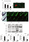
EP2 signalling mediates chronic inflammation in cerebral aneurysm walls. (A) Effects of EP2 deficiency on macrophage infiltration in cerebral aneurysm walls. The number of CD68-positive macrophages was determined in immunostained sections. The number of animals is shown in parentheses under each bar. Data represent mean ± SEM. **P < 0.01. Representative images are shown in the right panel. Bar = 20 µm. (B) Immunostaining for pro-inflammatory mediators in walls of cerebral aneurysms in littermates of Ptger2 heterozygous (Ptger2+/−) and Ptger2-deficient (Ptger2−/−) mice. Black arrows in Elastica van Gieson (EvG) staining (left) indicate the centre of the cerebral aneurysm or its corresponding position. White arrows indicate the internal elastic lamina of arterial walls. Bar = 20 µm. p-p65: phosphorylated NF-κB p65 subunit. (C) Effect of EP2 deficiency on NF-κB activation and expression of pro-inflammatory mediators in cerebral arteries. Representative images of Western blot analyses are shown in the upper panels and the quantification by densitometric analysis is shown in the lower panel (n = 5). α-Tubulin was used as an internal control. 0 or 5 M: before and at 5 months after induction of cerebral aneurysm. Data represent mean ± SEM. *P < 0.05.
Figure 4
Figure 4
Induction of CCL2 expression by EP2 stimulation via NF-κB pathway in ECs. (A) Effects of EP agonists on the mRNA level of CCL2 in primary ECs from human carotid artery (EP1 agonist; ONO-D1-004, EP2 agonist; ONO-AE1-259, EP3 agonist; ONO-AE-248, EP4 agonist; ONO-AE1-329). After 24 h stimulation with the respective receptor agonists (0.5 µM), mRNA levels of CCL2 were determined by RT-PCR (n = 6). Data represent mean ± SEM. *P < 0.05, **P < 0.01. (B) Effects of ONO-AE1-259, an EP2 agonist, on phosphorylation of p65 subunit of NF-κB (p-p65) and CCL2 protein. The left panel shows representative images of Western blot analyses. Middle and right panels show the quantification by the densitometric analyses of phospho-p65 signals (middle) and CCL2 signals (right), respectively (n = 5). Data represent mean ± s,e,mean. *P < 0.05. **P < 0.01. (C) Effects of Ptger2 depletion by RNAi on NF-κB phosphorylation and CCL2 expression induced by ONO-AE1-259. The left panel shows representative phosphorylated p65 subunit (p-p65) and EP2 signals in Western blot analyses under the indicated conditions. The middle panel shows quantification by the densitometric analyses of phosphorylated-p65 signals (n = 5). The right panel shows mRNA levels of CCL2 from ECs pretreated with siRNA for Ptger2 or control siRNA without or with subsequent EP2 stimulation (n = 6). Data represent mean ± SEM. **P < 0.01. (D) Effect of NF-κB inhibition by decoy oligodeoxynucleotide on EP2-induced CCL2 expression (n = 6). Data represent mean ± SEM. *P < 0.05, **P < 0.01.
Figure 5
Figure 5
A proposed model for the formation of cerebral aneurysm induced by shear stress.

References

    1. Alexander SPH, Mathie A, Peters JA. Guide to receptors and channels (GRAC), 4th edition (2009) Br J Pharmacol. 2009;158(Suppl 1):S1–S254. - PMC - PubMed
    1. Alnaes MS, Isaksen J, Mardal KA, Romner B, Morgan MK, Ingebrigtsen T. Computation of hemodynamics in the circle of Willis. Stroke. 2007;38:2500–2505. - PubMed
    1. Aoki T, Nishimura M. Targeting chronic inflammation in cerebral aneurysms: focusing on NF-κB as a putative target of medical therapy. Expert Opin Ther Targets. 2010;14:265–273. - PubMed
    1. Aoki T, Kataoka H, Morimoto M, Nozaki K, Hashimoto N. Macrophage-derived matrix metalloproteinase-2 and -9 promote the progression of cerebral aneurysms in rats. Stroke. 2007a;38:162–169. - PubMed
    1. Aoki T, Kataoka H, Shimamura M, Nakagami H, Wakayama K, Moriwaki T, et al. NF-κB is a key mediator of cerebral aneurysm formation. Circulation. 2007b;116:2830–2840. - PubMed

Publication types

MeSH terms