Review
. 2011 Jun 23;117(25):6768-76.
doi: 10.1182/blood-2011-02-334623. Epub 2011 Mar 22.

Thymic T-cell development in allogeneic stem cell transplantation

Affiliations
Review

Thymic T-cell development in allogeneic stem cell transplantation

Werner Krenger et al. Blood. .

Abstract

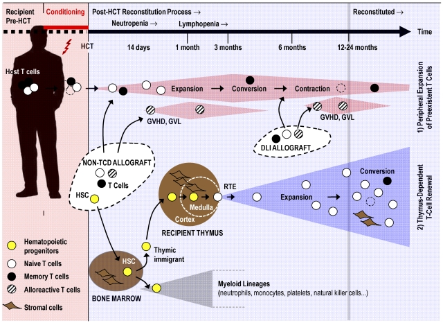
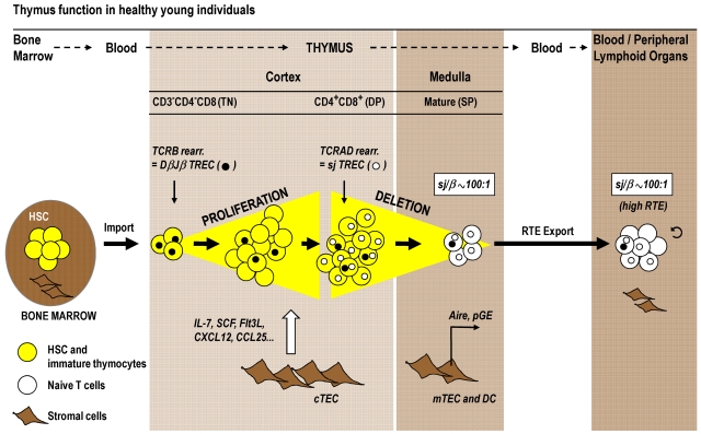
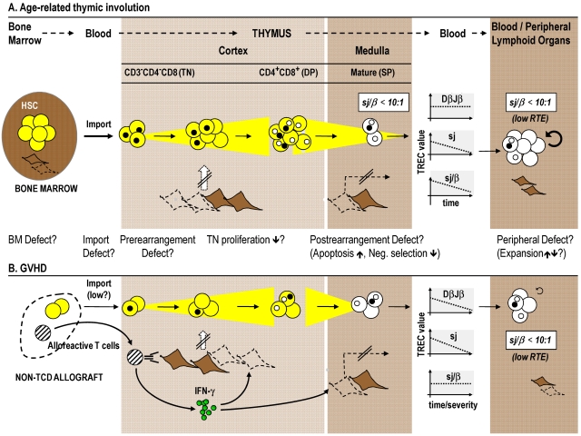
Cytoreductive conditioning regimens used in the context of allogeneic hematopoietic cell transplantation (HCT) elicit deficits in innate and adaptive immunity, which predispose patients to infections. As such, transplantation outcomes depend vitally on the successful reconstruction of immune competence. Restoration of a normal peripheral T-cell pool after HCT is a slow process that requires the de novo production of naive T cells in a functionally competent thymus. However, there are several challenges to this regenerative process. Most notably, advanced age, the cytotoxic pretransplantation conditioning, and posttransplantation alloreactivity are risk factors for T-cell immune deficiency as they independently interfere with normal thymus function. Here, we discuss preclinical allogeneic HCT models and clinical observations that have contributed to a better understanding of the transplant-related thymic dysfunction. The identification of the cellular and molecular mechanisms that control regular thymopoiesis but are altered in HCT patients is expected to provide the basis for new therapies that improve the regeneration of the adaptive immune system, especially with functionally competent, naive T cells.


Figures

Figure 1
Figure 1
T-cell regenerative pathways after allogeneic HCT. Pretransplantation conditioning reduces the patient's existing naive (○) and memory (●) T cells. Post-HCT regeneration of peripheral T cells is accomplished by 2 mechanisms: (1) Cell numbers of residual host T cells and mature donor T cells transferred via a non-TCD stem cell allograft or via DLI expand initially in response to homeostatic signals or cognate antigen. Alloreactive donor T cells (which can mediate GVHD, graft-versus-leukemia, or both; dashed symbols) contribute also to the thymus-independent cell compartment. This T-cell pool is altered as a result of memory T-cell conversion, skewing of the TCR repertoire, and pool size contraction (broken lines indicate senescent T cells). (2) New naive T cells are generated from hematopoietic progenitors (yellow symbols) in a thymus-dependent regenerative mechanism. The stromal cells of the thymus (thymic medullary and cortical compartments are separated from each other by a dashed line) support the differentiation of descendants of thymic immigrants into mature T lymphocytes. Recent thymic emigrants (RTEs) expand in the periphery in response to homeostatic signals and after stimulation by their cognate antigen. Under physiologic conditions, the thymic export assures a constant and life-long supply with naive T cells harboring a diverse TCR repertoire. After allogeneic HCT, the thymus-dependent T-cell renewal is a slow process as it can take up to 12 to 24 months to be completed under favorable conditions. However, advanced age, conditioning, and GVHD impair thymus function and thus interfere with naive T-cell regeneration. The putative mechanisms are discussed in Figure 3.
Figure 2
Figure 2
Normal thymic T-cell maturation and export. T-cell development requires the import of a bone marrow-derived progenitor population (yellow symbols) via the blood circulation into the thymus cortex. During optimal thymus function in healthy young persons, TN cells proliferate strongly in the cortex in response to signals provided by cTECs (eg, interleukin-7, stem cell factor, chemokines). The subsequent DP stage of thymocyte maturation is subject to thymic-positive selection. In parallel to their migration to the medulla, DP cells differentiate into SP thymocytes, which are subjected to the negative selection process. pGE in Aire-expressing mTECs plays a crucial role in clonal deletion. The mature T cells (white symbols) are exported as RTE via blood circulation to peripheral lymphoid tissues. The magnitude of thymic export can be assessed by TREC analysis. This approach is based on the fact that thymocytes undergo 2 sequential rounds of TCR rearrangements that form 2 families of TREC as byproducts. TCRB rearrangement in TN cells forms several DβJβ TREC species (●), which are diluted among the expanding cell population before the TCRAD is rearranged in DP cells, which generates sjTREC (○). Thymic export can be measured via determination of the sj/β ratio. This signature for RTE indicates the extent of TN cell proliferation, which is in turn a key determinant for the extent of thymic export. The sj/β ratio remains independently of peripheral cell division at approximately 100:1 in young persons. DC indicates dendritic cells; and sj/β, calculated ratio between sjTREC and DβJβTREC.
Figure 3
Figure 3
The effects of age and GVHD on T-cell maturation and export. Age-related changes and GVHD affect T-cell development in prethymic, thymic, and post-thymic compartments. (A) Thymus function itself peaks in the first and second decades of life but then declines in the course of the involution process. During senescence, the sjTREC frequencies decline, whereas DβJβ species remain unchanged. The resultant decrease in the sj/β ratio (which falls below a value of 1:10) indicates lower TN proliferation, which hence explains the decline in RTE export in aged persons. (B) GVHD interference with intrathymic maturation is indicated by parallel decreases in DβJβTREC and sjTREC frequencies. The result is an unchanged sj/β ratio in peripheral blood T cells, which implies that thymopoiesis is affected either before TCRB chain rearrangement and TN proliferation and/or at a time subsequent to TCRAD rearrangement (eg, apoptosis of postrearrangement thymocytes). Acute GVHD therefore impairs thymic function independently from age. Both age-related thymus involution and GVHD are characterized by changes in the thymic stromal microenvironment. Lack of proper stromal function consequently leads to impairment of thymocyte development. In preclinical allogeneic HCT models, the demise of TECs (which may include the death of tolerance-inducing mTECs that express Aire) is linked to alloreactive donor T cells secreting intrathymically IFN-γ in response to activation by host TEC. Abbreviations and symbols are the same as in Figure 2. Broken lines indicate stromal cell death; and strikethrough arrows, deficient TEC function. BM indicates bone marrow.

References

    1. Gratwohl A, Baldomero H. Trends of hematopoietic stem cell transplantation in the third millennium. Curr Opin Hematol. 2009;16(6):420–426. - PubMed
    1. Ballen KK, King RJ, Chitphakdithai P, et al. The national marrow donor program: 20 years of unrelated donor hematopoietic cell transplantation. Biol Blood Marrow Transplant. 2008;14(9 suppl):2–7. - PubMed
    1. Gooley TA, Chien JW, Pergam SA, et al. Reduced mortality after allogeneic hematopoietic-cell transplantation. N Engl J Med. 2010;363(22):2091–2101. - PMC - PubMed
    1. Deeg HJ, Sandmaier BM. Who is fit for allogeneic transplantation? Blood. 2010;116(23):4762–4770. - PMC - PubMed
    1. Pasquini MC, Wang Z. Current use and outcome of hematopoietic stem cell transplantation: II. CIBMTR Summary Slides 2009. CIBMTR Newsletter. 2009;15(2):7–11.

Publication types

MeSH terms