. 2011 Jun;90(6):810-6.
doi: 10.1177/0022034511402995. Epub 2011 Mar 22.

Bisphosphonates induce senescence in normal human oral keratinocytes

Affiliations

Bisphosphonates induce senescence in normal human oral keratinocytes

R H Kim et al. J Dent Res. 2011 Jun.

Abstract

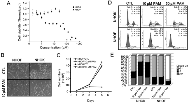
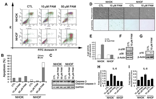
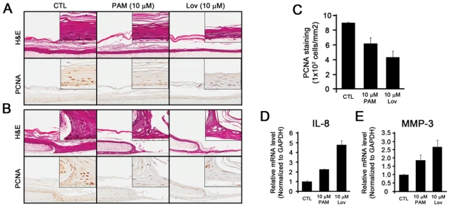
Bisphosphonate-related osteonecrosis of the jaw (BRONJ) commonly occurs in individuals receiving bisphosphonates (BPs) with clinical manifestations of the exposed necrotic bone. Although defective wound healing of soft tissue is frequently, if not always, observed in BRONJ, the effects of BPs on oral soft tissue or cells remain unknown. To investigate the effects of BPs on cells of oral mucosal tissue, we studied the effect of pamidronate (PAM), one of the BPs most commonly administered to cancer patients, on the phenotypes of normal human oral keratinocytes (NHOK) and fibroblasts (NHOF). When exposed to PAM at 10 µM, NHOK, not NHOF, underwent senescence: NHOK overexpressed senescence-associated β-galactosidase (SA-β-Gal), p16INK4A, IL-6, and IL-8. When exposed to a higher level (50 µM) of PAM, NHOK maintained senescent phenotypes, but NHOF underwent apoptosis. PAM-induced senescence in NHOK is mediated, in part, via geranylgeranylation of the mevalonate pathway. Our in vitro 3D oral mucosal tissue construction studies further demonstrated that PAM induced senescence and impaired re-epithelialization of oral mucosa. Analysis of these data indicates that premature senescence of oral mucosal cells and subsequent defective soft-tissue wound healing might be partly responsible for the development of BRONJ in individuals receiving PAM or other BPs.


Figures

Figure 1.
Figure 1.
Pamidronate induces cytotoxic effects on oral mucosal cells in a cell-type-specific manner. (A) NHOK and NHOF were treated with various doses of pamidronate (PAM). Four days after treatment, cells were subjected to MTT assay. Experiments were performed in quadruplicate, and bars indicate standard errors. (B) NHOK and NHOF were treated with 10 µM PAM for 4 days, and cells were photographed at 100X. (C) NHOK and NHOF were plated onto the 6-well plates 24 hrs before cells were treated with 10 µM PAM. Every 2 days, cells were either harvested for counting or changed with fresh medium containing 10 µM PAM. (D) NHOK and NHOF were treated with 10 and 50 µM PAM for 4 days, and cells were harvested, stained with PI, and subjected to flow cytometry analysis. (E) The percentages of cell cycle contents in Fig. 1D were quantitated and are presented with the bar graph.
Figure 2.
Figure 2.
Pamidronate induces senescence, but not apoptosis, in NHOK. (A) NHOK and NHOF were treated with 10 and 50 µM PAM for 4 days, stained with FITC Annexin V and PI, and subjected to Flow Cytometry analysis. (B) The percentages of cells in early and late apoptosis in Fig. 2A were quantitated and are presented in the bar graph. (C) NHOK and NHOF were treated with 10 and 50 µM for 4 days, and cells were harvested for Western blot analysis against cleaved Caspase-3. GAPDH was also probed for a loading control. (D) NHOK and NHOF were treated with 10 µM PAM for 4 days and stained for SA-β-Gal activity. The photographs were taken at 100X. (E) β-Gal-positive NHOK and NHOF were quantitated by use of the MarkerGene Cellular Senescence Microtiterplate Assay Kit. (F) PAM-treated NHOK were subjected to Western blotting against phosphorylated p38 and total p38. β-actin was probed for the loading control. (G) Western blotting against p16 was also probed. (H, I) NHOK and NHOF treated with 10 or 50 µM PAM were harvested, mRNA was isolated, and cDNA was synthesized. qRT-PCR was run with primers for IL-6 and IL-8. Samples were run in triplicate and normalized to β-actin.
Figure 3.
Figure 3.
Geranylgerniol (GGOH), but not farnesol (FOH), partially reverses the effects of pamidronate in NHOK. (A) NHOK were treated with the indicated combinations (10 µM PAM, 1 µM FOH, 0.5 µM GGOH, 1 µM FOH/0.5 µM GGOH) for 6 days. Typical photographs were taken (100x). (B) NHOK were seeded at 2 × 10⁴ cells per well and treated with the indicated combinations of PAM, FOH, GGOH, or FOH/GGOH at day 1. The numbers were counted after 3 and 6 days. The experiment was performed in triplicate, with bars representing standard errors. (C) NHOK were plated at 2 × 10⁴ cells per well and treated with the indicated combinations of PAM, FOH, GGOH, or FOH/GGOH at day 1. After 6 days, cells were subjected to SA-β-gal activity. Cells in 3 independent fields were counted. Error bars represent standard errors.
Figure 4.
Figure 4.
Pamidronate attenuates the re-epithelialization of oral mucosal cells. A 3D oral mucosal wound-healing model was established by the organotypic raft culture system. (A) Five days after air-lifting of the transwell, the oral mucosal tissue constructs were fed with medium only (CTL), medium containing 10 µM PAM, or medium containing 10 µM lovastatin (Lov). The oral tissue constructs were harvested 2 wks after the air-lifting, and subjected to H&E staining (top panel) and IHC staining against IgG (1:200, not shown) and PCNA (1:200) (bottom panel). (B) The circular wound was created 1 wk after the air-lifting. The oral tissue constructs were harvested after 7 days and subjected to H&E staining (top panel) and IHC staining against PCNA (bottom panel). (C) Three independent fields were randomly selected, and PCNA-positive cells were counted per mm2. The bar represents the standard deviation. (D, E) The epithelial cells of the oral mucosal tissue constructs were dissected by LCM. Total RNAs were isolated, cDNAs were synthesized, and qRT-PCR was performed for the expression of IL-8 (D) and MMP-3 (E). The experiments were performed in triplicate, and values were normalized to GAPDH.

References

    1. Adams PD. (2009). Healing and hurting: molecular mechanisms, functions, and pathologies of cellular senescence. Mol Cell 36:2-14 - PubMed
    1. Adornato MC, Morcos I, Rozanski J. (2007). The treatment of BP associated osteonecrosis of the jaws with bone resection and autologous platelet-derived growth factors. J Am Dent Assoc 138:971-977 - PubMed
    1. Agis H, Blei J, Watzek G, Gruber R. (2010). Is zoledronate toxic to human periodontal fibroblasts? J Dent Res 89:40-45 - PubMed
    1. Chaturvedi V, Qin JZ, Denning MF, Choubey D, Diaz MO, Nickoloff BJ. (1999). Apoptosis in proliferating, senescent, and immortalized keratinocytes. J Biol Chem 274:23358-23367 - PubMed
    1. Chaturvedi V, Qin JZ, Stennett L, Choubey D, Nickoloff BJ. (2004). Resistance to UV-induced apoptosis in human keratinocytes during accelerated senescence is associated with functional inactivation of p53. J Cell Physiol 198:100-109 - PubMed

Publication types

LinkOut - more resources